Clinical Assessment & Protocol
Typical Presentation (HPI)
EN: Fever, headache, neck stiffness, and photophobia. AR: حمى، صداع، تيبس في الرقبة، ورهاب الضوء.
General Examination
EN: Positive Kernig and Brudzinski signs. AR: علامات كيرنيج وبرودزينسكي إيجابية.
Treatment Protocol
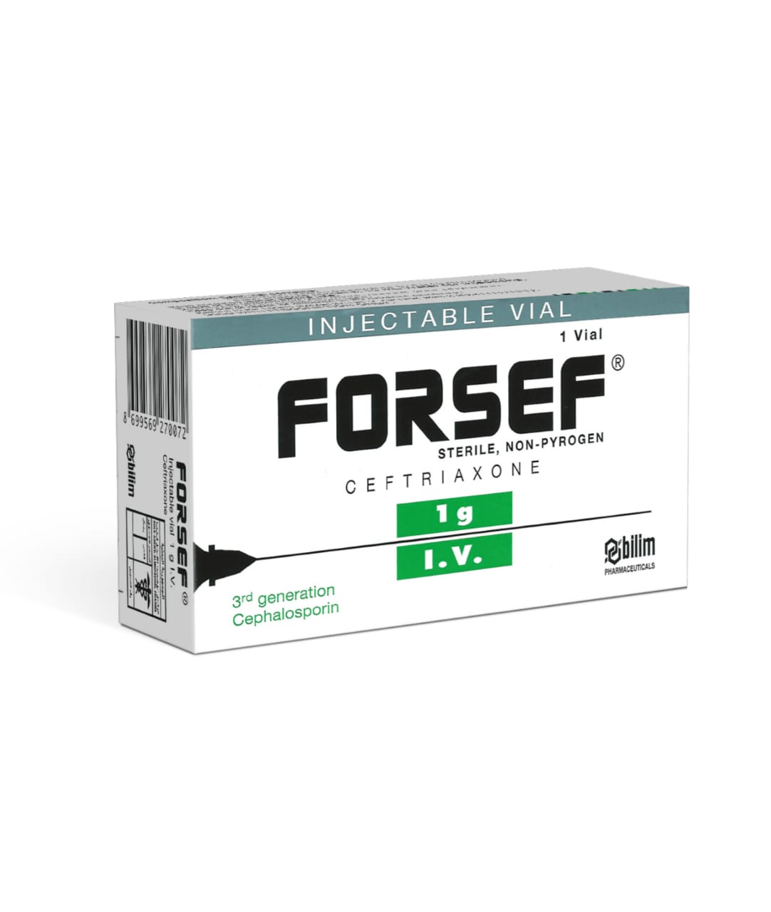
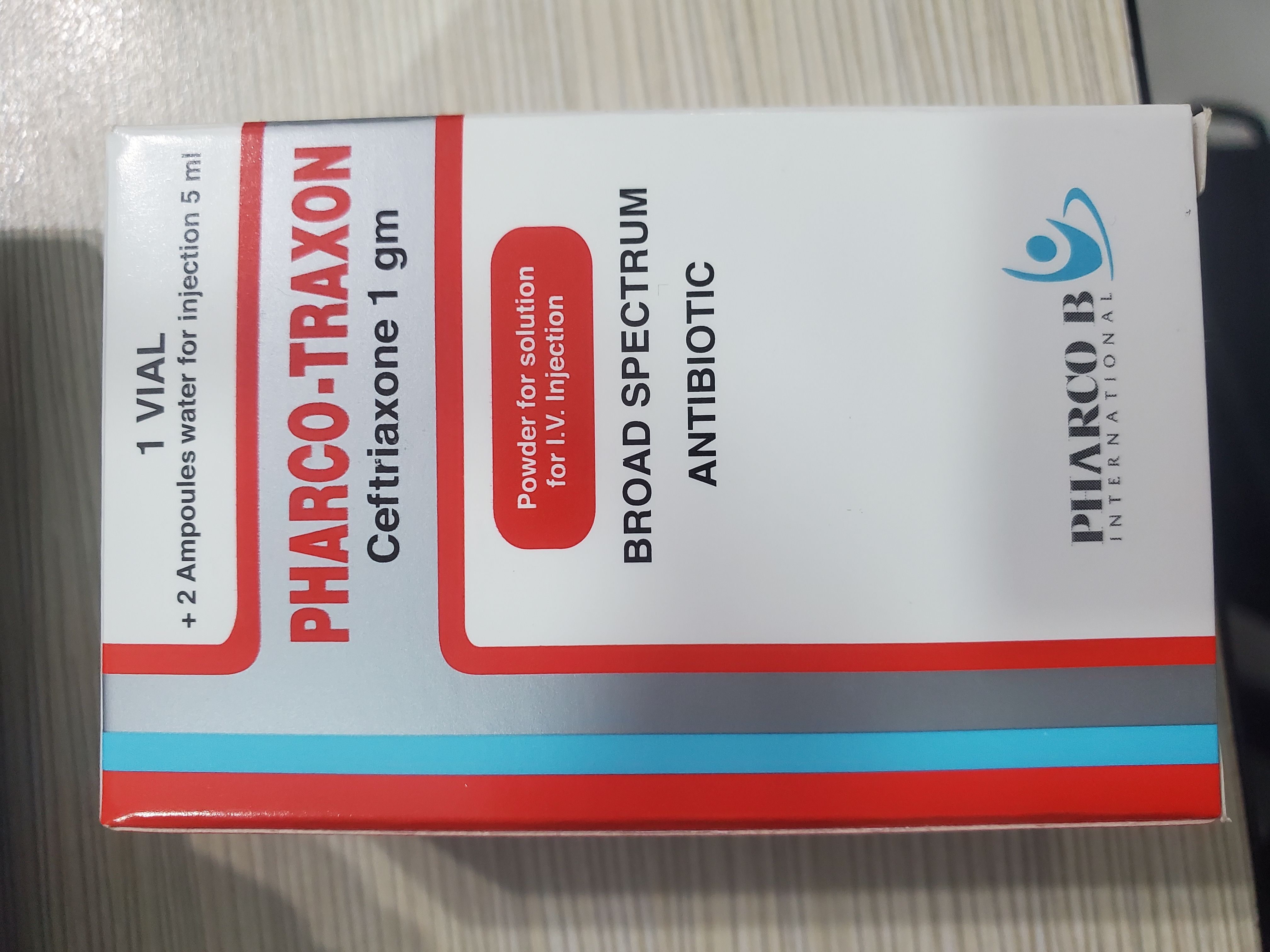
EN: Empiric IV antibiotics (ceftriaxone/vancomycin) and dexamethasone. AR: مضادات حيوية وريدية تجريبية (سيفترياكسون/فانكومايسين) وديكساميثازون.
Patient Education
EN: Explain the urgency and the need for close monitoring. AR: شرح الحالة الطارئة وضرورة المراقبة اللصيقة.
Systemic & Specialized Examinations
EN: S1, S2 present. No murmurs. AR: صوتا القلب الأول والثاني طبيعيان. لا توجد نفخات.
EN: Lungs clear to auscultation. AR: الرئتان صافيتان عند التسمع.
EN: Abdomen soft, non-tender. AR: البطن لين ولا يوجد ألم.
EN: Alert, oriented x3. No focal deficits. AR: المريض واعي ومدرك. لا يوجد عجز عصبي بؤري.
EN: Unremarkable or not routinely indicated. AR: طبيعي أو غير مطلوب روتينياً.
EN: Unremarkable or not routinely indicated. AR: طبيعي أو غير مطلوب روتينياً.
EN: Unremarkable or not routinely indicated. AR: طبيعي أو غير مطلوب روتينياً.
EN: Unremarkable or not routinely indicated. AR: طبيعي أو غير مطلوب روتينياً.
EN: Unremarkable or not routinely indicated. AR: طبيعي أو غير مطلوب روتينياً.
Orthopedic & Trauma Assessments
EN: Unremarkable or not routinely indicated. AR: طبيعي أو غير مطلوب روتينياً.
EN: Unremarkable or not routinely indicated. AR: طبيعي أو غير مطلوب روتينياً.
Clinical Guide: Pediatric Bacterial Meningitis
1. Comprehensive Introduction & Overview
Pediatric bacterial meningitis represents one of the most critical medical emergencies in clinical pediatrics. It is defined as an acute, purulent infection of the subarachnoid space, involving the leptomeninges (the arachnoid and pia mater) and the underlying cerebrospinal fluid (CSF). Unlike viral meningitis, which is typically self-limiting, bacterial meningitis is characterized by rapid progression, high morbidity, and significant mortality if not addressed with aggressive, targeted clinical intervention.
In the pediatric population, the disease burden varies significantly by age, immunization status, and geographic region. Despite the global implementation of conjugate vaccines (such as the Haemophilus influenzae type b (Hib), Streptococcus pneumoniae, and Neisseria meningitidis vaccines), bacterial meningitis remains a leading cause of long-term neurological sequelae, including hearing loss, cognitive impairment, and epilepsy.
2. Technical Specifications: Etiology and Pathophysiology
Etiology by Age Group
The causative organisms of pediatric bacterial meningitis are highly age-dependent, reflecting the host's immune maturity and exposure risks.
| Age Group | Common Pathogens |
|---|---|
| Neonates (0–28 days) | Streptococcus agalactiae (Group B Strep), Escherichia coli, Listeria monocytogenes |
| Infants (1–3 months) | S. agalactiae, E. coli, S. pneumoniae, N. meningitidis |
| Children (>3 months) | S. pneumoniae, N. meningitidis |
Pathophysiology: The Cascade of Infection
The development of bacterial meningitis occurs through a complex multi-step mechanism:
- Colonization and Invasion: Pathogens colonize the nasopharyngeal mucosa. Through the secretion of IgA proteases, bacteria evade mucosal immunity and enter the bloodstream.
- Bacteremia: The bacteria survive within the intravascular space, resisting complement-mediated killing and phagocytosis via polysaccharide capsules.
- Blood-Brain Barrier (BBB) Crossing: Bacteria cross the BBB, likely via infected endothelial cells or by "Trojan horse" mechanisms involving infected leukocytes.
- Subarachnoid Space Proliferation: Once in the CSF, the lack of effective host immune factors (low complement and antibody levels) allows for rapid bacterial replication.
- Inflammatory Cascade: Bacterial cell wall components (teichoic acid, lipopolysaccharide) trigger the release of pro-inflammatory cytokines (TNF-α, IL-1β). This results in:
- Vasogenic Edema: Increased permeability of the BBB.
- Cytotoxic Edema: Cellular swelling due to metabolic failure.
- Interstitial Edema: Obstructive hydrocephalus resulting from impaired CSF resorption.
3. Clinical Indications, Presentation, and Staging
Clinical Presentation
The presentation of bacterial meningitis is highly variable. In neonates and young infants, symptoms are often non-specific, necessitating a high index of suspicion.
- Classic Triad (Often absent in children): Fever, nuchal rigidity, and altered mental status.
- Neonatal Presentation: Temperature instability, lethargy, poor feeding, bulging fontanelle, seizures, and respiratory distress.
- Older Pediatric Presentation: Photophobia, phonophobia, vomiting, headache, and signs of meningeal irritation (Kernig’s and Brudzinski’s signs).
Clinical Staging/Grading
While there is no universally standardized "staging" system like cancer, clinicians utilize the Meningitis Severity Score or clinical assessment of neurological status (GCS) to guide management.
| Stage | Clinical Indicators |
|---|---|
| Early/Mild | Irritability, low-grade fever, no focal neurological deficits. |
| Moderate | Persistent vomiting, moderate lethargy, meningismus present. |
| Severe | Coma, seizures, signs of increased intracranial pressure (ICP), shock. |
Differential Diagnosis
It is imperative to differentiate bacterial meningitis from other pathologies:
* Viral Meningoencephalitis: Typically presents with a milder course; CSF shows lymphocytic pleocytosis.
* Brain Abscess: Focal neurological deficits are more prominent; imaging (MRI) is diagnostic.
* Diabetic Ketoacidosis (DKA): Can present with altered mental status and vomiting.
* Intracranial Hemorrhage: Sudden onset of focal signs and meningeal irritation.
4. Key Diagnostic Tests
Diagnostic protocols must be rapid and structured. Time is brain.
Lumbar Puncture (LP)
The gold standard for diagnosis. CSF analysis typically reveals:
* Appearance: Cloudy or turbid.
* WBC Count: Usually >1,000 cells/µL with neutrophil predominance.
* Glucose: Decreased (<40 mg/dL or <40% of blood glucose).
* Protein: Elevated (>100 mg/dL).
* Gram Stain/Culture: Essential for targeted antibiotic therapy.
Ancillary Testing
- Blood Cultures: Positive in 50–80% of cases.
- PCR/Multiplex Panels: Rapid identification of common pathogens, especially if antibiotics were started before the LP.
- Neuroimaging (CT/MRI): Indicated only if there are signs of increased ICP, focal deficits, or prolonged seizures to rule out hydrocephalus or abscess before LP.
5. Risks, Side Effects, and Contraindications
Risks of Intervention
- Lumbar Puncture: Risk of herniation (rare, if ICP is significantly elevated), spinal headache, or localized infection.
- Antibiotic Therapy: Risk of hypersensitivity reactions, secondary C. difficile infection, or nephrotoxicity (if aminoglycosides are used).
- Dexamethasone Therapy: Used to reduce inflammatory sequelae; must be administered before or concurrently with the first dose of antibiotics. If given after, efficacy is significantly reduced.
Contraindications to Immediate LP
LP should be delayed in the presence of:
1. Hemodynamic instability.
2. Active seizures.
3. Signs of impending brain herniation (cranial nerve palsies, fixed pupils, decerebrate posturing).
4. Local skin infection at the site of puncture.
6. Long-Term Prognosis and Sequelae
The prognosis is contingent upon the speed of diagnosis and the specific pathogen. Neurological sequelae occur in roughly 10–20% of survivors.
- Sensorineural Hearing Loss: The most common complication, particularly with S. pneumoniae.
- Cognitive Deficits: Learning disabilities, intellectual impairment, and attention deficits.
- Motor Impairment: Spasticity, paresis, or ataxia.
- Epilepsy: Chronic seizure disorders resulting from cortical scarring.
7. Frequently Asked Questions (FAQ)
1. Is bacterial meningitis contagious?
Yes. N. meningitidis and H. influenzae are transmissible via respiratory droplets. Prophylactic antibiotics are often required for close household contacts.
2. Can my child get meningitis even if they are vaccinated?
Vaccines are highly effective, but not 100%. Breakthrough infections can occur, although they are generally less severe.
3. What is the role of steroids in treatment?
Dexamethasone is used to modulate the inflammatory response in the subarachnoid space, which helps reduce the risk of hearing loss and other neurological complications.
4. How long does the child need to be in the hospital?
Typically, a minimum of 7 to 14 days of intravenous antibiotics is required, depending on the causative pathogen and clinical response.
5. What are the signs of increased intracranial pressure?
Persistent vomiting, bradycardia, hypertension, irregular breathing (Cushing’s triad), and bulging fontanelle in infants.
6. Will my child need a follow-up hearing test?
Yes. Universal auditory screening is mandatory for all pediatric survivors of bacterial meningitis shortly after recovery.
7. What is the difference between meningitis and encephalitis?
Meningitis is an inflammation of the protective membranes covering the brain; encephalitis is an inflammation of the brain tissue itself.
8. Can I perform an LP if the child is on antibiotics?
Yes, but the yield of the culture may be reduced. PCR testing is highly recommended in "partially treated" cases.
9. What is the most common cause of bacterial meningitis in a 6-month-old?
S. pneumoniae remains the most common etiology in this age group in most developed nations.
10. How quickly does bacterial meningitis progress?
It can progress from mild symptoms to critical illness within hours. It is considered a medical emergency requiring immediate stabilization.
8. Clinical Management Summary Table
| Management Pillar | Action Item |
|---|---|
| Stabilization | ABCs (Airway, Breathing, Circulation), IV access, fluid resuscitation. |
| Empiric Therapy | Ceftriaxone + Vancomycin (add Ampicillin for neonates or if Listeria suspected). |
| Adjunctive Therapy | Dexamethasone (0.15 mg/kg Q6H for 4 days). |
| Monitoring | Frequent neurological checks, seizure precautions, strict intake/output. |
| Prevention | Ensure completion of Hib, PCV13/15/20, and Meningococcal vaccination series. |
Disclaimer: This guide is intended for educational and clinical reference purposes for healthcare professionals. It does not replace institutional protocols or individual clinical judgment. Always consult current IDSA (Infectious Diseases Society of America) guidelines for specific dosing and regional resistance patterns.
Related Clinical Integration
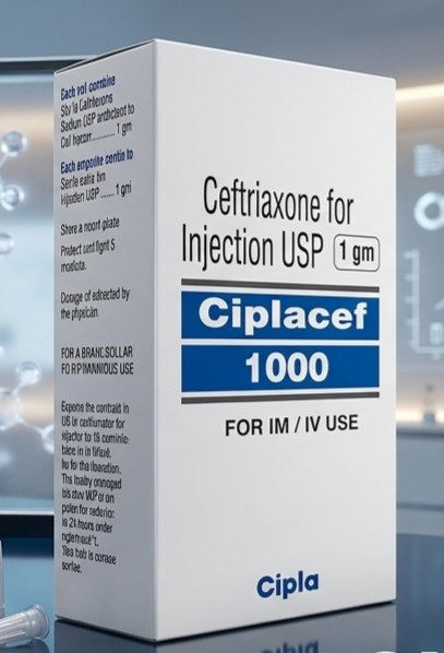
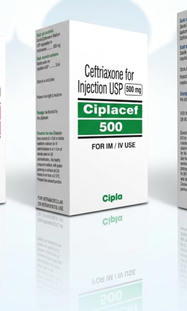
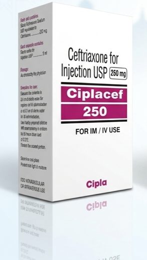
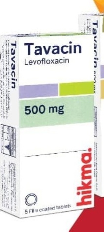
In a modern clinical setting, the management of pediatric bacterial meningitis requires a multidisciplinary approach centered on rapid diagnostic precision and aggressive therapeutic intervention. Upon clinical suspicion, immediate diagnostic protocols must be initiated, including Blood Cultures and Cranial imaging (MRI/CT) to assess for complications such as increased intracranial pressure. The cornerstone of treatment involves the urgent Intravenous antibiotic administration of empiric agents, most commonly Ceftriaxone / سيفترياكسون 1 g, Ceftriaxone for Injection / سيفترياكسون للحقن 500 mg, or Vancomycin / فانكومايسين 1g, to ensure adequate cerebrospinal fluid penetration. In critically ill patients, supportive care may necessitate Oxygen Administration, Fluid resuscitation, and Nutritional Support (TPN/Enteral) to maintain metabolic stability. Furthermore, if the patient presents with signs of sepsis-induced organ dysfunction, clinicians may need to monitor Serum electrolyte monitoring and Renal function testing to guide potential escalation to [Renal replacement therapy (e.g., hemodialysis, continuous venovenous hemodialysis)](https://yemenhealthos.com/ar
Treatment & Management Options
Recommended Medications