Clinical Assessment & Protocol
Typical Presentation (HPI)
EN: Patient presents with acute onset of localized mid-lumbar back pain following [mechanism of injury, e.g., mechanical fall/lifting]. Pain is described as sharp, non-radiating, and exacerbated by movement, standing, or spinal loading. Patient denies bowel/bladder incontinence, saddle anesthesia, or progressive lower extremity weakness. AR: يعاني المريض من ألم حاد وموضعي في أسفل الظهر بعد [آلية الإصابة، مثل: سقوط أو رفع ثقل]. يصف المريض الألم بأنه حاد، غير منتشر، ويزداد سوءاً مع الحركة أو الوقوف أو تحميل الوزن على العمود الفقري. ينفي المريض وجود سلس بولي أو برازي، أو خدر في منطقة السرج، أو ضعف متزايد في الأطراف السفلية.
General Examination
EN: Spine: Focal midline tenderness to palpation at the L1 vertebral level. No step-off deformity noted. Range of motion is severely limited by pain in all planes. Neurological: Motor strength 5/5 in bilateral lower extremities. Sensation intact to light touch in all dermatomes. Deep tendon reflexes 2+ and symmetric. Negative straight leg raise test bilaterally. No signs of myelopathy or cauda equina syndrome. AR: العمود الفقري: وجود ألم موضعي عند الجس في خط المنتصف عند مستوى الفقرة القطنية الأولى (L1). لا توجد تشوهات أو انزياحات ملموسة. نطاق الحركة محدود بشدة بسبب الألم في جميع الاتجاهات. الفحص العصبي: القوة العضلية 5/5 في الأطراف السفلية. الإحساس سليم للمس الخفيف في جميع القطاعات الجلدية. المنعكسات الوترية العميقة 2+ ومتناظرة. اختبار رفع الساق المستقيمة سلبي على الجانبين. لا توجد علامات لاعتلال النخاع أو متلازمة ذيل الفرس.
Treatment Protocol
EN: Conservative management initiated: Activity modification with avoidance of heavy lifting and spinal flexion. Pain control via scheduled NSAIDs and/or acetaminophen. Orthotic bracing (TLSO) recommended for stabilization and pain relief. Referral to physical therapy for core stabilization once acute phase subsides. Follow-up imaging in 2-4 weeks to monitor for progressive collapse. AR: البدء بالعلاج التحفظي: تعديل النشاط مع تجنب رفع الأثقال وثني العمود الفقري. السيطرة على الألم باستخدام مضادات الالتهاب غير الستيرويدية و/أو الباراسيتامول بانتظام. يوصى باستخدام دعامة الظهر (TLSO) للتثبيت وتخفيف الألم. إحالة إلى العلاج الطبيعي لتقوية عضلات الجذع بمجرد زوال المرحلة الحادة. إجراء تصوير متابعة خلال 2-4 أسابيع لمراقبة أي انهيار إضافي للفقرة.
Patient Education
EN: You have been diagnosed with a stable compression fracture of the L1 vertebra. Healing typically takes 6-12 weeks. Maintain a neutral spine posture; avoid bending, twisting, or lifting objects heavier than 5 lbs. Wear your prescribed brace as directed. Seek immediate emergency care if you experience sudden loss of bowel/bladder control, numbness in the groin area, or new weakness in your legs. AR: تم تشخيصك بكسر انضغاطي مستقر في الفقرة القطنية الأولى (L1). يستغرق الشفاء عادةً من 6 إلى 12 أسبوعاً. حافظ على وضعية مستقيمة للعمود الفقري؛ تجنب الانحناء أو الالتواء أو رفع أجسام أثقل من 2 كجم. ارتدِ الدعامة الموصوفة لك حسب التعليمات. اطلب الرعاية الطارئة فوراً إذا شعرت بفقدان مفاجئ للتحكم في البول أو البراز، أو خدر في منطقة العجان، أو ضعف جديد في ساقيك.
Systemic & Specialized Examinations
EN: Distinct radiculopathy (L4/L5/S1). Strict Cauda Equina precautions documented. AR: اعتلال عصبي جذري واضح. تم توثيق تحذيرات متلازمة ذيل الفرس بصرامة.
Orthopedic & Trauma Assessments
EN: Heavy lifting incident with spinal rotation, or insidious degenerative disc disease. AR: حادث رفع أوزان ثقيلة مع دوران للعمود الفقري، أو انزلاق غضروفي تدريجي.
EN: Antalgic gait. Exhibits a 'list' (sciatic scoliosis) away from the affected side. Difficulty with heel/toe walk. AR: مشية متألمة. يظهر ميلاً (جنف وركي) لتخفيف الضغط. صعوبة في المشي على الكعب/الأصابع.
EN: Loss of normal lumbar lordosis. Severe paraspinal muscle spasm. AR: فقدان التقوس القطني الطبيعي. تشنج عضلي شديد حول الفقرات.
EN: Straight Leg Raise (SLR): Strongly positive at 30-45°. Slump test positive. AR: اختبار رفع الساق المستقيمة (SLR): إيجابي بقوة عند 30-45 درجة.
EN: Weakness (4/5) in EHL (L5) or Plantarflexion (S1). AR: ضعف (4/5) في باسطة الإبهام (L5) أو الثني الأخمصي (S1).
EN: Hypoesthesia to pinprick over the foot dorsum (L5) or lateral border (S1). AR: نقص الإحساس للوخز على ظهر القدم (L5) أو الجانب الوحشي (S1).
EN: Achilles (S1) diminished 1+. Patellar (L4) 2+. AR: منعكس وتر أخيل ضعيف 1+. منعكس الرضفة طبيعي 2+.
EN: DP and PT pulses 2+ symmetric. AR: النبضات الطرفية طبيعية.
Clinical Comprehensive Guide: Vertebral Compression Fracture (VCF), Lumbar, L1, Initial, Closed
1. Comprehensive Introduction & Overview
A Vertebral Compression Fracture (VCF) at the L1 level represents one of the most common skeletal injuries encountered in clinical orthopedics and emergency medicine. Defined as the collapse of a vertebral body, the L1 vertebra serves as a critical junction point between the thoracic spine (rigid) and the lumbar spine (mobile), making it a frequent site of mechanical failure.
In the context of an "Initial, Closed" diagnosis, the patient is presenting with a fresh injury where the integrity of the surrounding skin and soft tissue remains intact, and no protrusion of bone fragments into the spinal canal has occurred (neurologically intact). This guide serves as a definitive clinical resource for medical professionals to understand the lifecycle of an L1 VCF, from acute presentation to long-term management.
2. Deep-Dive: Pathophysiology and Biomechanics
The L1 vertebra is subject to high axial loading forces. When the force applied to the vertebral body exceeds the structural capacity of the trabecular bone, a fracture occurs.
The Mechanism of Failure
- Axial Compression: The superior endplate is driven into the vertebral body.
- Flexion-Compression: Often associated with "wedge" fractures, where the anterior portion of the vertebral body loses height while the posterior wall remains relatively stable.
- Bone Density Influence: In patients with underlying osteoporosis, the structural failure occurs at a much lower energy threshold compared to healthy, high-density bone.
Pathophysiological Stages
| Stage | Description |
|---|---|
| Acute (0-2 weeks) | Edema within the vertebral marrow; micro-fractures of trabeculae; pain from periosteal stretching. |
| Sub-acute (2-8 weeks) | Initiation of callus formation; stabilization of the fracture fragment. |
| Chronic (>12 weeks) | Remodeling phase; potential for pseudoarthrosis if healing is incomplete. |
3. Clinical Indications and Standard Presentation
Clinical Symptoms
Patients presenting with an initial, closed L1 VCF typically report:
* Localized Mid-Back Pain: Pain specifically centered at the thoracolumbar junction.
* Radicular Symptoms: While often absent in "closed" fractures, some patients may report referred pain around the flank or groin.
* Postural Changes: Increased kyphosis (hunching) if the fracture is significant.
* Functional Limitation: Severe difficulty with transitions (sitting to standing) or ambulation.
Physical Examination Findings
- Palpation: Point tenderness over the L1 spinous process.
- Percussion: Increased pain with gentle percussion over the L1 level.
- Neurological Screen: Critical to rule out cord or nerve root compression. Test dermatomes (L1-S1), myotomes (hip flexion, knee extension, dorsiflexion), and deep tendon reflexes.
- Gait Analysis: Antalgic gait or compensatory trunk leaning.
4. Differential Diagnosis
Distinguishing an L1 VCF from other pathologies is essential for appropriate care:
- Pathologic Fracture: Must rule out metastatic disease (multiple myeloma, lung, breast, or prostate cancer) using MRI.
- Disc Herniation: Usually presents with more pronounced radiculopathy.
- Spondylolisthesis: Often shows a "step-off" deformity on palpation.
- Musculoligamentous Strain: Typically resolves within 2 weeks; lacks the radiographic evidence of vertebral height loss.
5. Diagnostic Testing Protocols
For an initial L1 VCF, the following diagnostic hierarchy is standard:
Imaging Modalities
- Radiography (X-Ray): AP and Lateral views. Look for "wedge" deformity, loss of vertebral height (>20%), or cortical disruption.
- Computed Tomography (CT): Gold standard for assessing bony integrity, retropulsion of fragments, and the "three-column" stability (Denis classification).
- Magnetic Resonance Imaging (MRI): Essential for identifying "acute" versus "chronic" fractures via STIR (Short Tau Inversion Recovery) sequencing. Acute fractures will show marrow edema.
6. Risks, Side Effects, and Contraindications
Risks of Non-Management
- Progressive Kyphosis: Leads to chronic back pain and restrictive lung disease due to chest wall compression.
- Pseudoarthrosis: Failure of the bone to unite, leading to permanent instability.
- Neurological Deficit: Delayed onset of nerve root impingement if the fracture progresses.
Contraindications for Conservative Management
- Neurological Compromise: Any evidence of cauda equina symptoms or weakness.
- Instability: Fracture involving the posterior column (e.g., burst fracture) requires surgical consultation.
- Intractable Pain: Failure to respond to analgesics, warranting vertebral augmentation (Kyphoplasty/Vertebroplasty).
7. Clinical Staging and Prognosis
Prognostic Indicators
- Bone Density: Patients with T-scores below -2.5 (osteoporosis) have a significantly higher risk of subsequent fractures.
- Fracture Severity: A loss of >50% of vertebral height is a strong predictor of future deformity.
- Age: Advanced age correlates with slower healing and higher risk of secondary complications (e.g., pneumonia, DVT).
Long-Term Outlook
Most closed L1 VCFs resolve with conservative management (bracing, NSAIDs, physical therapy) within 8 to 12 weeks. However, the "cascade effect" must be monitored, as one vertebral fracture increases the risk of a secondary fracture by fivefold within the first year.
8. Massive FAQ Section
Q1: What does "Closed" mean in this diagnosis?
"Closed" indicates that the skin and surrounding soft tissues are intact, and the fracture has not resulted in a compound injury or exposed bone.
Q2: Is an L1 fracture considered "serious"?
Yes, as the L1 is the transition point of the spine. While many are managed non-surgically, they require careful monitoring to prevent progressive deformity.
Q3: How long until I can walk normally again?
Typically, patients can ambulate with a brace within days of the injury, but full functional recovery often takes 6-12 weeks.
Q4: Do I need surgery for an L1 VCF?
Not always. Surgery (Kyphoplasty or instrumentation) is reserved for cases with severe pain, neurological deficits, or significant structural instability.
Q5: What is the role of a TLSO brace?
A Thoracolumbar Sacral Orthosis (TLSO) brace provides external stabilization, reduces motion at the fracture site, and helps manage pain during the acute healing phase.
Q6: Can osteoporosis cause this without a fall?
Yes. "Fragility fractures" can occur during routine daily activities like coughing, sneezing, or lifting a light object in patients with severe osteoporosis.
Q7: Why is an MRI necessary if the X-ray shows the fracture?
An X-ray shows the bone structure, but an MRI shows the "age" of the fracture by detecting marrow edema, which is critical for determining if the pain is truly from the new injury.
Q8: What are the warning signs of nerve damage?
Seek immediate emergency care if you experience bowel or bladder incontinence, saddle anesthesia (numbness in the groin area), or sudden, progressive leg weakness.
Q9: Will I always have a hunchback (kyphosis)?
Minor height loss is common, but significant deformity can be mitigated with proper bracing, physical therapy, and osteoporosis treatment.
Q10: What should I avoid during the healing process?
Avoid heavy lifting, twisting of the spine, and high-impact activities for at least 3 months, or until cleared by your orthopedic specialist.
9. Clinical Management Summary Table
| Management Aspect | Recommendation |
|---|---|
| Analgesia | Acetaminophen/NSAIDs; consider short-term opioids if pain is severe. |
| Bracing | TLSO brace for 6-8 weeks for pain management and support. |
| Activity | "Activity as tolerated"—avoid bed rest, which increases risk of DVT. |
| Bone Health | Initiate Calcium/Vitamin D; evaluate for Bisphosphonate therapy. |
| Follow-up | Repeat X-rays at 6 weeks to monitor for further collapse. |
10. Conclusion for the Practitioner
The management of an "Initial, Closed L1 Vertebral Compression Fracture" requires a systematic approach that balances pain management with functional restoration. By utilizing high-quality diagnostic imaging and a structured follow-up plan, clinicians can prevent the long-term complications of kyphosis and chronic pain. The cornerstone of success lies in the early identification of patients at high risk for secondary fractures and the aggressive management of any underlying metabolic bone disease. Always prioritize the neurological exam, as the L1 level is the termination point of the spinal cord (conus medullaris), making it a high-stakes zone for clinical vigilance.
Related Clinical Integration
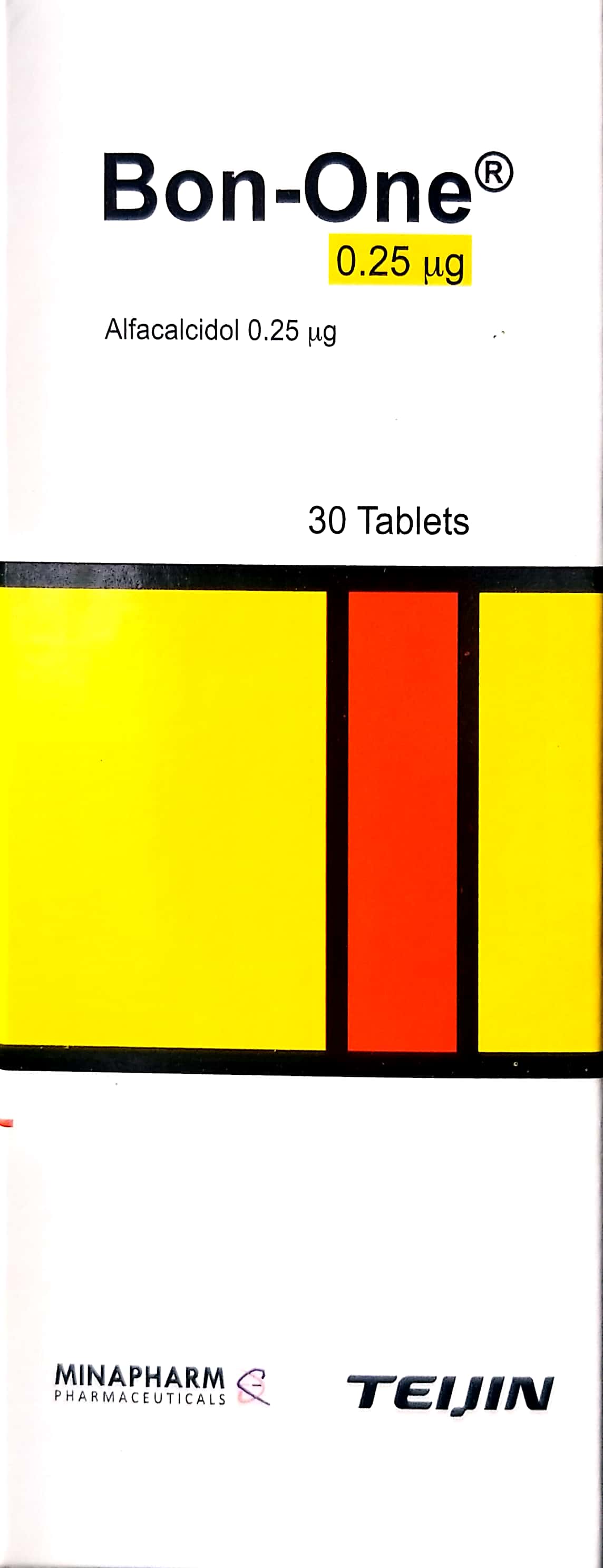
In the management of an L1 vertebral compression fracture, a multidisciplinary approach is essential to address both acute structural instability and underlying bone health. Clinical intervention often begins with pharmacological stabilization using agents such as Bon-one / بون-ون 0.25mcg, Calcitonin Nasal Spray / بخاخ الكالسيتونين الأنفي 200 IU/spray, or Forteo / فورتيو 20mcg/dose to improve bone density and manage pain. For patients presenting with refractory pain or significant deformity, surgical stabilization via Kyphoplasty / رأب الحدباء (عملية كبرى في غرف العمليات) or, in specific cases of malignancy, Kyphoplasty/Vertebroplasty for Pathologic Oncologic Fracture / رأب الحدب/رأب الفقرات لكسر مرضي ورمي (عملية صغرى في العيادة) may be indicated. To ensure evidence-based decision-making, clinicians should consult comprehensive resources such as Mastering the Management of Thoracic and Lumbosacral Fractures and review specialized board-level literature, including AAOS & ABOS Spine Surgery MCQs (Set 4): Spinal Trauma, Cervical Myelopathy & Adult Scoliosis, [AAOS & ABOS Spine Surgery MCQs (Set 4): Vertebral Fractures & Adult Deformity | 2000 Board Review](https://www.hutaifortho.com/en/hub/spine-surgery-2009-set-4-mcqs-405