Clinical Assessment & Protocol
Typical Presentation (HPI)
EN: Fatigue, malar rash, joint pain, and unexplained fever. AR: إرهاق، طفح جلدي على شكل فراشة، ألم مفصلي، وحمى غير مبررة.
General Examination
EN: Malar rash, alopecia, oral ulcers, and joint tenderness. AR: طفح الفراشة، ثعلبة، تقرحات فموية، وإيلام مفصلي.
Treatment Protocol
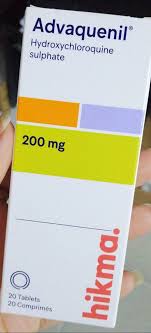
EN: Hydroxychloroquine, corticosteroids, and immunosuppressants. AR: هيدروكسي كلوروكين، كورتيكوستيرويدات، ومثبطات المناعة.
Patient Education
EN: Sun protection, medication adherence, and early reporting of fever. AR: الحماية من الشمس، الالتزام بالأدوية، والإبلاغ المبكر عن الحمى.
Systemic & Specialized Examinations
EN: S1, S2 present. No murmurs. AR: صوتا القلب الأول والثاني طبيعيان. لا توجد نفخات.
EN: Lungs clear to auscultation. AR: الرئتان صافيتان عند التسمع.
EN: Abdomen soft, non-tender. AR: البطن لين ولا يوجد ألم.
EN: Alert, oriented x3. No focal deficits. AR: المريض واعي ومدرك. لا يوجد عجز عصبي بؤري.
EN: Unremarkable or not routinely indicated. AR: طبيعي أو غير مطلوب روتينياً.
EN: Unremarkable or not routinely indicated. AR: طبيعي أو غير مطلوب روتينياً.
EN: Unremarkable or not routinely indicated. AR: طبيعي أو غير مطلوب روتينياً.
EN: Unremarkable or not routinely indicated. AR: طبيعي أو غير مطلوب روتينياً.
EN: Unremarkable or not routinely indicated. AR: طبيعي أو غير مطلوب روتينياً.
Orthopedic & Trauma Assessments
EN: Unremarkable. Not routinely indicated or affected by this specific systemic pathology. AR: طبيعي. غير مطلوب روتينياً أو غير متأثر بهذا المرض الجهازي.
EN: Unremarkable. Not routinely indicated or affected by this specific systemic pathology. AR: طبيعي. غير مطلوب روتينياً أو غير متأثر بهذا المرض الجهازي.
EN: Unremarkable or not routinely indicated. AR: طبيعي أو غير مطلوب روتينياً.
EN: Unremarkable or not routinely indicated. AR: طبيعي أو غير مطلوب روتينياً.
EN: Unremarkable. Not routinely indicated or affected by this specific systemic pathology. AR: طبيعي. غير مطلوب روتينياً أو غير متأثر بهذا المرض الجهازي.
EN: Unremarkable. Not routinely indicated or affected by this specific systemic pathology. AR: طبيعي. غير مطلوب روتينياً أو غير متأثر بهذا المرض الجهازي.
EN: Unremarkable. Not routinely indicated or affected by this specific systemic pathology. AR: طبيعي. غير مطلوب روتينياً أو غير متأثر بهذا المرض الجهازي.
EN: Unremarkable. Not routinely indicated or affected by this specific systemic pathology. AR: طبيعي. غير مطلوب روتينياً أو غير متأثر بهذا المرض الجهازي.
EN: Unremarkable. Not routinely indicated or affected by this specific systemic pathology. AR: طبيعي. غير مطلوب روتينياً أو غير متأثر بهذا المرض الجهازي.
Comprehensive Clinical Guide: Systemic Lupus Erythematosus (SLE)
Systemic Lupus Erythematosus (SLE) represents the prototype of systemic autoimmune diseases. Characterized by a profound loss of self-tolerance, it manifests as a multi-system inflammatory disorder mediated by autoantibodies and immune complex deposition. As a chronic, relapsing-remitting condition, it poses significant diagnostic and therapeutic challenges to clinicians across rheumatology, nephrology, and internal medicine.
1. Clinical Definition and Overview
Systemic Lupus Erythematosus is a chronic inflammatory disease of autoimmune origin, characterized by the production of a wide array of autoantibodies against nuclear, cytoplasmic, and cell-surface antigens. The clinical spectrum is vast, ranging from mild mucocutaneous involvement to life-threatening glomerulonephritis, neuropsychiatric manifestations, and hematologic cytopenias.
Key Epidemiological Characteristics
- Predominance: Strong female predilection (9:1 ratio in reproductive years).
- Age of Onset: Typically between 15 and 45 years.
- Genetic Susceptibility: Association with HLA-DR2 and HLA-DR3 alleles; deficiencies in early complement components (C1q, C2, C4).
2. Etiology and Pathophysiology: The Molecular Mechanism
The pathogenesis of SLE is multifactorial, involving an intricate interplay between genetic predisposition, hormonal factors, and environmental triggers.
The Mechanism of Immune Dysregulation
- Impaired Clearance of Apoptotic Debris: Defects in the clearance of apoptotic cells result in the accumulation of nuclear antigens.
- B-Cell Hyperactivity: Loss of peripheral tolerance leads to the activation of autoreactive B-cells.
- Type I Interferon (IFN) Signature: A hallmark of SLE, the "interferon signature" involves the chronic overexpression of Type I IFNs, which promotes dendritic cell maturation and T-cell activation.
- Immune Complex Deposition: The formation of antigen-antibody complexes leads to the activation of the classical complement pathway, resulting in tissue-specific inflammation via Type III hypersensitivity reactions.
Environmental Triggers
- Ultraviolet (UV) Radiation: Induces keratinocyte apoptosis and DNA damage.
- Infectious Agents: Epstein-Barr virus (EBV) molecular mimicry.
- Hormonal Influence: Estrogen exacerbates the immune response, while androgens appear protective.
3. Clinical Presentation and Staging
SLE is known as "The Great Imitator" due to its protean manifestations. Diagnosis relies on the 2019 EULAR/ACR Classification Criteria.
Common Clinical Manifestations
| System | Common Findings |
|---|---|
| Constitutional | Fatigue, fever, weight loss, lymphadenopathy. |
| Mucocutaneous | Malar rash, discoid rash, photosensitivity, oral ulcers. |
| Musculoskeletal | Non-erosive arthritis, arthralgia, Jaccoud’s arthropathy. |
| Renal | Lupus Nephritis (Proteinuria, hematuria, casts). |
| Hematologic | Leukopenia, lymphopenia, thrombocytopenia, hemolytic anemia. |
| Neuropsychiatric | Seizures, psychosis, cognitive dysfunction, transverse myelitis. |
Staging: The Lupus Nephritis (LN) Classification (ISN/RPS)
Renal involvement is a primary determinant of prognosis, categorized as follows:
* Class I: Minimal mesangial LN.
* Class II: Mesangial proliferative LN.
* Class III: Focal LN.
* Class IV: Diffuse LN (The most severe, requiring aggressive immunosuppression).
* Class V: Membranous LN.
* Class VI: Advanced sclerosing LN.
4. Diagnostic Approach and Key Investigations
Diagnosis is rarely based on a single test. It requires a synthesis of clinical suspicion and serological support.
Essential Diagnostic Tests
- Antinuclear Antibody (ANA): Highly sensitive (95-99%) but lacks specificity.
- Anti-dsDNA: Highly specific for SLE; titers often correlate with disease activity and renal involvement.
- Anti-Smith (Sm): Pathognomonic for SLE; highly specific.
- Antiphospholipid Antibodies (aPL): Includes Lupus Anticoagulant, Anticardiolipin, and Anti-beta2-glycoprotein I; associated with thrombotic risk.
- Complement Levels (C3, C4): Low levels indicate active disease (consumption during immune complex formation).
Differential Diagnosis
Clinicians must distinguish SLE from:
* Mixed Connective Tissue Disease (MCTD).
* Rheumatoid Arthritis (RA).
* Sjögren’s Syndrome.
* Drug-Induced Lupus (commonly due to hydralazine, procainamide, isoniazid).
* Fibromyalgia (often mimics the diffuse pain of SLE).
5. Risks, Contraindications, and Therapeutic Management
Management Pillars
- Hydroxychloroquine (HCQ): The cornerstone of therapy for all SLE patients; reduces flares and improves long-term survival.
- Glucocorticoids: Used for induction of remission; requires careful tapering to avoid long-term toxicity.
- Immunosuppressants: Mycophenolate Mofetil (MMF) or Cyclophosphamide for severe organ-threatening disease.
- Biologics: Belimumab (BLyS inhibitor) and Anifrolumab (Type I IFN receptor antagonist).
Contraindications/Warnings
- Pregnancy: Certain medications (e.g., Mycophenolate, Cyclophosphamide) are teratogenic.
- Live Vaccines: Generally contraindicated in patients on high-dose immunosuppression.
- Cardiovascular Risk: SLE is an independent risk factor for premature atherosclerosis.
6. Long-Term Prognosis
The prognosis of SLE has improved dramatically over the last 50 years, with 10-year survival rates now exceeding 90%. However, morbidity remains high due to:
* Chronic Organ Damage: Specifically renal failure and cardiovascular disease.
* Infections: Often secondary to chronic immunosuppressive therapy.
* Malignancy: Increased risk of certain lymphomas and HPV-related cervical dysplasia.
7. Frequently Asked Questions (FAQ)
1. Is SLE considered a hereditary disease?
While it has a genetic component, it is not strictly hereditary. Having a first-degree relative with SLE increases risk, but the majority of patients do not have a family history.
2. Why is sun protection so important for SLE patients?
UV light can trigger both cutaneous flares and systemic activity by causing apoptosis of skin cells, which releases autoantigens that stimulate the immune system.
3. Can a person with SLE have a healthy pregnancy?
Yes. However, it is considered a high-risk pregnancy. It requires careful planning, medication adjustment (e.g., stopping teratogens), and monitoring for antiphospholipid syndrome.
4. What is the difference between "Lupus" and "Lupus Nephritis"?
Lupus is the systemic disease. Lupus Nephritis is a specific, severe manifestation where the kidneys are targeted by immune complexes.
5. Do all SLE patients have a positive ANA test?
Approximately 95% do. A patient with a negative ANA is highly unlikely to have SLE, though "seronegative" cases exist.
6. What is the role of Hydroxychloroquine?
It is the only medication proven to reduce mortality in SLE, manage skin/joint symptoms, and prevent future organ damage. It is recommended for almost all patients regardless of severity.
7. How often should I monitor my blood work?
During active disease, monthly monitoring may be necessary. In stable, quiescent disease, labs are typically checked every 3–6 months.
8. What is the "Lupus Fog"?
It refers to cognitive dysfunction, including memory loss, trouble concentrating, and confusion, which is a common and debilitating neuropsychiatric symptom of SLE.
9. Are there foods that can cure or trigger Lupus?
There is no "Lupus diet." However, an anti-inflammatory diet (low in processed foods, high in Omega-3s) can help manage general health. Avoid alfalfa sprouts, which contain L-canavanine, known to trigger flares in some patients.
10. Is there a cure for SLE?
Currently, there is no cure. However, with modern therapeutic advances, the majority of patients can achieve "clinical remission" or "low disease activity," allowing for a normal life expectancy and quality of life.
Disclaimer
This guide is for educational and informational purposes only and does not constitute medical advice, diagnosis, or treatment. Always seek the advice of a board-certified rheumatologist or qualified health provider regarding any medical condition.