Clinical Assessment & Protocol
Typical Presentation (HPI)
EN: Patient with known Systemic Lupus Erythematosus (SLE) presents for follow-up of cryoglobulinemia. Reports [symptoms, e.g., arthralgia, purpura, or fatigue] persisting for [duration]. Current disease activity is [stable/worsening]. AR: مريض مشخص بالذئبة الحمراء الجهازية (SLE) يراجع للمتابعة بخصوص وجود الغلوبولينات البردية (cryoglobulinemia). يشكو من [الأعراض، مثل آلام المفاصل، فرفرية، أو إرهاق] منذ [المدة]. نشاط المرض الحالي [مستقر/متدهور].
General Examination
EN: Patient appears [well/ill-appearing], alert and oriented x3. Vital signs: BP [value], HR [value], Temp [value]. No acute distress noted. AR: المريض يبدو [بحالة جيدة/مريضاً]، واعٍ ومدرك للزمان والمكان. العلامات الحيوية: ضغط الدم [القيمة]، نبض القلب [القيمة]، درجة الحرارة [القيمة]. لا توجد علامات ضيق حاد.
Treatment Protocol
EN: Plan includes: 1. Continue current SLE therapy: [medication]. 2. Manage cryoglobulinemia with [treatment, e.g., immunosuppressants/plasmapheresis]. 3. Monitor [labs, e.g., C3, C4, cryoglobulin levels] in [timeframe]. AR: الخطة العلاجية تشمل: 1. الاستمرار على علاج الذئبة الحالي: [الدواء]. 2. تدبير الغلوبولينات البردية بـ [العلاج، مثل مثبطات المناعة/فصادة البلازما]. 3. مراقبة [التحاليل، مثل C3, C4، مستويات الغلوبولينات البردية] خلال [الفترة الزمنية].
Patient Education
EN: Discussed the association between SLE and cryoglobulinemia. Advised patient to avoid cold exposure, monitor for new skin rashes or joint pain, and adhere strictly to medication regimen. AR: تمت مناقشة العلاقة بين الذئبة الحمراء والغلوبولينات البردية. تم نصح المريض بتجنب التعرض للبرد، ومراقبة أي طفح جلدي جديد أو آلام في المفاصل، والالتزام الصارم بالخطة العلاجية.
Systemic & Specialized Examinations
EN: Skin examination reveals [presence/absence] of palpable purpura, livedo reticularis, or ulcers, specifically on [location]. AR: فحص الجلد يظهر [وجود/غياب] فرفرية مجسوسة، تزرّق شبكي، أو تقرحات، خاصة في [الموقع].
Orthopedic & Trauma Assessments
EN: Peripheral pulses are [symmetrical/asymmetrical] and [strong/diminished] in [location, e.g., lower extremities], consistent with potential vasculitic involvement. AR: النبضات المحيطية [متناظرة/غير متناظرة] و[قوية/ضعيفة] في [الموقع، مثل الأطراف السفلية]، بما يتوافق مع احتمالية وجود إصابة وعائية.
Medical Guide: Systemic Lupus Erythematosus (associated with cryoglobulinemia)
1. Comprehensive Introduction & Overview
Systemic Lupus Erythematosus (SLE) is a chronic, multifactorial autoimmune disease characterized by the production of autoantibodies and immune complex formation, leading to widespread inflammation and damage across various organ systems. It is a complex disorder with a highly variable clinical presentation, ranging from mild skin and joint involvement to severe, life-threatening organ damage affecting the kidneys, brain, heart, and lungs.
Cryoglobulinemia, on the other hand, is a condition defined by the presence of cryoglobulins in the blood. Cryoglobulins are immunoglobulins (antibodies) or immunoglobulin complexes that reversibly precipitate at temperatures below normal body temperature (typically 4°C) and redissolve upon rewarming. This temperature-dependent precipitation can lead to vascular occlusion and inflammation, particularly in small and medium-sized blood vessels, resulting in a distinct set of clinical manifestations known as cryoglobulinemic vasculitis.
The association between SLE and cryoglobulinemia represents a significant clinical entity. While cryoglobulinemia is most commonly associated with chronic infections (especially Hepatitis C virus, HCV) or lymphoproliferative disorders, it can also occur as a secondary phenomenon in autoimmune diseases like SLE. When cryoglobulinemia complicates SLE, it typically manifests as Type II or Type III mixed cryoglobulinemia. This co-occurrence often indicates a more severe disease phenotype, characterized by heightened immune activity and specific organ involvement, such as glomerulonephritis and peripheral neuropathy, which can significantly impact patient morbidity and mortality. Understanding this specific association is crucial for accurate diagnosis, targeted management, and improved patient outcomes.
2. Deep-Dive into Technical Specifications / Mechanisms: Etiology and Pathophysiology
The interplay between SLE and cryoglobulinemia involves complex immunological mechanisms rooted in both conditions' etiologies and pathophysiologies.
Etiology of Systemic Lupus Erythematosus (SLE)
The precise cause of SLE remains unknown, but it is understood to arise from a complex interaction of:
* Genetic Predisposition: A strong genetic component is evident, with numerous susceptibility genes identified (e.g., HLA-DRB1, STAT4, IRF5, PTPN22). These genes influence immune regulation, antigen presentation, and cytokine signaling.
* Environmental Triggers: Exposure to ultraviolet (UV) light, certain drugs (e.g., procainamide, hydralazine), viral infections (e.g., Epstein-Barr virus), and cigarette smoking can initiate or exacerbate SLE.
* Hormonal Factors: Estrogen is thought to play a role, given the higher prevalence of SLE in women, particularly during reproductive years. Androgens may have protective effects.
* Epigenetic Modifications: Alterations in DNA methylation and histone modification can influence gene expression, contributing to immune dysregulation.
Pathophysiology of SLE
SLE's pathophysiology is characterized by a breakdown of immune tolerance, leading to:
1. Aberrant Apoptosis & Impaired Clearance of Apoptotic Debris: Increased cell death and defective clearance of cellular remnants expose nuclear and cytoplasmic autoantigens.
2. Autoantibody Production: The immune system mistakenly recognizes self-antigens as foreign, leading to the production of a vast array of autoantibodies (e.g., antinuclear antibodies [ANA], anti-double-stranded DNA [anti-dsDNA], anti-Sm, anti-RNP).
3. Immune Complex Formation: Autoantibodies bind to self-antigens, forming circulating immune complexes.
4. Immune Complex Deposition: These complexes deposit in various tissues (e.g., kidneys, skin, joints, blood vessels), activating the complement system and recruiting inflammatory cells.
5. Inflammation and Tissue Damage: Complement activation, cytokine release (e.g., type I interferons, TNF-α), and cellular infiltration (e.g., macrophages, neutrophils) drive chronic inflammation, leading to organ damage and dysfunction.
Etiology of Cryoglobulinemia in the Context of SLE
When cryoglobulinemia occurs in SLE, it is almost exclusively Type II (monoclonal IgM-RF with polyclonal IgG) or Type III (polyclonal IgM-RF with polyclonal IgG) mixed cryoglobulinemia.
* Polyclonal B-cell Activation: SLE is characterized by chronic B-cell hyperactivity and dysregulation, leading to the production of a wide range of autoantibodies, including rheumatoid factor (RF). This sustained polyclonal activation can directly contribute to the formation of Type III cryoglobulins.
* Immune Complex Composition: In mixed cryoglobulinemia, IgM-RF antibodies (often polyclonal in SLE) bind to the Fc portion of IgG molecules. These IgM-IgG complexes can then precipitate in the cold.
* Secondary Cryoglobulinemia: Unlike essential mixed cryoglobulinemia (where no underlying cause is identified), cryoglobulinemia in SLE is secondary to the underlying autoimmune process. The chronic inflammatory state and immune dysregulation inherent to SLE create an environment conducive to cryoglobulin formation.
Pathophysiology of Cryoglobulinemia-Associated Complications
The presence of cryoglobulins leads to distinct pathological processes:
1. Cold-Induced Precipitation: In cooler parts of the body (e.g., extremities, skin capillaries), cryoglobulins precipitate, forming aggregates that can directly obstruct small blood vessels.
2. Cryoglobulinemic Vasculitis: The precipitated cryoglobulins act as immune complexes, depositing in vessel walls. This triggers complement activation (often leading to markedly low C4 levels) and recruits inflammatory cells, particularly neutrophils.
3. Leukocytoclastic Vasculitis: The characteristic histological finding is leukocytoclastic vasculitis, where neutrophils infiltrate and damage vessel walls, leading to fibrinoid necrosis and fragmentation of neutrophil nuclei (leukocytoclasis).
4. Ischemia and Organ Damage: Vascular occlusion and inflammatory damage to vessel walls result in tissue ischemia, purpura, skin ulcers, peripheral neuropathy, and specific forms of glomerulonephritis (e.g., membranoproliferative glomerulonephritis).
3. Extensive Clinical Indications & Usage
The clinical presentation of SLE associated with cryoglobulinemia is a blend of typical SLE features and specific manifestations driven by cryoglobulinemic vasculitis.
Standard Presentation
Patients typically present with:
A. General SLE Manifestations (consistent with standard SLE diagnosis criteria):
* Constitutional: Profound fatigue, unexplained fever, weight loss.
* Musculoskeletal: Arthralgia (joint pain) and non-erosive arthritis (most commonly in small joints of hands, wrists, knees), myalgia.
* Cutaneous: Malar rash (butterfly rash), discoid lupus rash, photosensitivity, oral/nasal ulcers.
* Serositis: Pleuritis (chest pain with breathing), pericarditis (chest pain).
* Renal: Lupus nephritis, manifesting as proteinuria, hematuria, hypertension, and potentially renal insufficiency.
* Neurological: Headaches, cognitive dysfunction, seizures, psychosis, stroke.
* Hematological: Anemia (hemolytic, chronic disease), leukopenia (lymphopenia), thrombocytopenia.
B. Cryoglobulinemia-Specific Manifestations (often superimposed or exacerbating SLE symptoms):
* Cutaneous Vasculitis:
* Palpable Purpura: The most common manifestation, typically on lower extremities, non-blanching red-purple spots or patches.
* Livedo Reticularis: Lacy, reticular pattern of reddish-blue discoloration of the skin, often cold-induced.
* Skin Ulcers/Necrosis: Particularly on distal extremities, fingers, and toes, due to severe vasculitis and ischemia.
* Raynaud's Phenomenon: Spasm of blood vessels in response to cold or stress, causing fingers and toes to turn white, then blue, then red.
* Peripheral Neuropathy:
* Sensory: Numbness, tingling, burning pain, often in a stocking-glove distribution.
* Motor: Weakness, muscle atrophy.
* Often a painful, distal, symmetrical polyneuropathy or mononeuritis multiplex.
* Renal Involvement:
* Cryoglobulinemic Glomerulonephritis: Can occur independently or alongside lupus nephritis. Often presents as membranoproliferative glomerulonephritis (MPGN) Type I or Type III, characterized by proteinuria, hematuria, and renal insufficiency. This form can be more aggressive than typical lupus nephritis.
* Arthralgia/Arthritis: Non-erosive inflammatory arthritis, which may be difficult to distinguish from typical lupus arthritis but can be more persistent.
* Gastrointestinal: Abdominal pain, nausea, vomiting, or diarrhea due to visceral vasculitis.
* Constitutional: Exacerbated fatigue, weakness, and malaise.
Clinical Staging/Grading & Disease Progression
There is no formal "staging" system for SLE associated with cryoglobulinemia. Instead, the condition is managed by:
* Assessing SLE Disease Activity: Using validated indices like SLEDAI (Systemic Lupus Erythemia Disease Activity Index), BILAG (British Isles Lupus Assessment Group), or ECLAM (European Consensus Lupus Activity Measure).
* Evaluating Organ Damage: Using indices like SLICC-ACR Damage Index.
* Characterizing Cryoglobulinemic Vasculitis: Documenting the extent and severity of cryoglobulin-related organ involvement (skin, nerves, kidneys).
* Renal Biopsy: Crucial for classifying glomerulonephritis (lupus nephritis class and presence of cryoglobulinemic features like intraluminal thrombi or MPGN pattern).
Disease progression is highly variable. The presence of cryoglobulinemia often implies a more active and severe disease course, particularly if there is significant renal or neurological involvement. Regular monitoring is essential to detect flares and manage complications.
Differential Diagnosis
Differentiating SLE with cryoglobulinemia from other conditions is critical:
* Primary (Essential) Mixed Cryoglobulinemia: Rule out other causes of cryoglobulinemia, particularly HCV infection, HIV, and lymphoproliferative disorders.
* Other Vasculitides: ANCA-associated vasculitis (GPA, MPA, EGPA), polyarteritis nodosa, IgA vasculitis (Henoch-Schönlein purpura).
* Other Autoimmune Diseases: Rheumatoid arthritis, Sjögren's syndrome (which can also be associated with cryoglobulinemia), scleroderma, inflammatory myopathies.
* Infections: Chronic bacterial endocarditis, Lyme disease, other viral infections.
* Hematological Malignancies: Lymphoma, multiple myeloma, Waldenström's macroglobulinemia (especially for Type I cryoglobulinemia).
* Drug-Induced Vasculitis: Certain medications can induce vasculitic syndromes.
Key Diagnostic Tests
A comprehensive diagnostic workup includes:
A. For Systemic Lupus Erythematosus:
* Autoantibody Panel:
* Antinuclear Antibodies (ANA): Highly sensitive (95-98%) for SLE, but not specific.
* Anti-double-stranded DNA (anti-dsDNA): Highly specific for SLE, correlates with disease activity, especially lupus nephritis.
* Anti-Sm: Highly specific for SLE.
* Anti-RNP, Anti-Ro (SSA), Anti-La (SSB): May be present.
* Complement Levels:
* C3 and C4: Often decreased in active SLE due to consumption by immune complexes. C4 is particularly low in mixed cryoglobulinemia.
* Inflammatory Markers: Erythrocyte Sedimentation Rate (ESR) and C-reactive protein (CRP) (CRP may be less elevated in SLE unless serositis or infection is present).
* Complete Blood Count (CBC): To detect cytopenias (anemia, leukopenia, lymphopenia, thrombocytopenia).
* Renal Function Tests: Serum creatinine, BUN, estimated GFR.
* Urinalysis with Urine Protein-to-Creatinine Ratio or 24-hour Urine Protein: To screen for and quantify proteinuria and hematuria, indicative of lupus nephritis.
B. For Cryoglobulinemia:
* Cryoglobulin Testing:
* Critical Sample Handling: Blood collected in a pre-warmed tube, transported immediately to the laboratory at 37°C, allowed to clot at 37°C, then serum separated. Serum is incubated at 4°C for up to 7 days, with daily inspection for precipitate formation.
* Cryocrit: Quantification of the cryoglobulin precipitate as a percentage of total serum volume.
* Cryoglobulin Typing (Immunofixation/Immunoelectrophoresis): To identify the immunoglobulin classes and light chain types present in the cryoprecipitate (e.g., IgM-kappa monoclonal with polyclonal IgG for Type II, or polyclonal IgM and IgG for Type III).
* Rheumatoid Factor (RF): Often positive, especially in Type II/III cryoglobulinemia.
* Complement Levels (C4): Markedly low C4 is a hallmark of mixed cryoglobulinemia due to classical pathway activation. C3 may also be low.
* Hepatitis C Virus (HCV) Serology and PCR: Essential to rule out HCV infection, as it is the most common cause of mixed cryoglobulinemia.
* Other Viral Markers: HIV, Hepatitis B.
* Serum Protein Electrophoresis (SPEP) and Immunofixation: To screen for monoclonal gammopathy, especially if Type I cryoglobulinemia is suspected (less common in SLE).
* Biopsies:
* Skin Biopsy: Of purpuric lesions, showing leukocytoclastic vasculitis with Ig and complement deposition.
* Kidney Biopsy: To assess for lupus nephritis class and specific cryoglobulinemic features (e.g., glomerular capillary thrombi, MPGN pattern).
* Nerve Biopsy: In cases of severe neuropathy, showing vasculitis or axonal degeneration.
4. Risks, Side Effects, or Contraindications (Complications and Prognosis)
The presence of cryoglobulinemia in SLE significantly influences the disease course, leading to specific complications and impacting long-term prognosis.
Major Complications of SLE with Cryoglobulinemia
- Severe Renal Disease: Cryoglobulinemic glomerulonephritis can be aggressive, leading to rapidly progressive renal failure, necessitating dialysis or kidney transplantation. It often presents as membranoproliferative glomerulonephritis, which can be resistant to standard lupus nephritis therapies.
- Peripheral Neuropathy: Can cause debilitating sensory and motor deficits, leading to chronic pain, numbness, weakness, and impaired mobility.
- Skin Ulcerations and Gangrene: Severe vasculitis can lead to extensive and painful skin ulcers, particularly on the lower extremities, which are prone to infection and may require surgical debridement or amputation in extreme cases.
- Gastrointestinal Vasculitis: Can result in ischemic bowel, perforations, or hemorrhage, leading to acute abdominal emergencies.
- Increased Risk of Infections: Both the disease itself and the immunosuppressive therapies used to treat it predispose patients to bacterial, viral, and fungal infections, which are a leading cause of mortality in SLE.
- Thrombotic Events: While less common than in some other cryoglobulinemias, vascular inflammation and immune complex deposition can potentially increase the risk of both arterial and venous thrombosis.
- Lymphoproliferative Disorders: Chronic B-cell stimulation and immune dysregulation, particularly in the context of Type II cryoglobulinemia, carry a small but increased long-term risk of developing B-cell lymphomas. This is a rare but serious complication.
- Cardiovascular Complications: Accelerated atherosclerosis is a known risk in SLE. Cryoglobulinemia-associated vasculitis can further contribute to vascular damage, potentially increasing cardiovascular risk.
Long-Term Prognosis
The long-term prognosis for patients with SLE associated with cryoglobulinemia is generally guarded and more challenging than for SLE alone.
* Increased Morbidity and Mortality: The presence of cryoglobulinemia, especially with significant renal or neurological involvement, is associated with increased morbidity and a higher risk of mortality compared to SLE patients without cryoglobulinemia.
* Organ Damage: Cumulative organ damage is often more pronounced due to the dual impact of SLE and cryoglobulinemic vasculitis.
* Treatment Resistance: Cryoglobulinemic manifestations can be more refractory to standard immunosuppressive therapies, often requiring more aggressive and specific treatments.
* Variable Course: The disease course is highly variable, with periods of remission and flares. Regular monitoring and proactive management are crucial.
* Impact on Quality of Life: Chronic pain, fatigue, organ damage, and treatment side effects can significantly impair the patient's quality of life.
Early diagnosis, aggressive immunosuppression targeting both SLE activity and cryoglobulinemic vasculitis, and meticulous management of complications are critical for improving outcomes.
5. Massive FAQ Section
Q1: What is the link between Systemic Lupus Erythematosus (SLE) and cryoglobulinemia?
A1: SLE is an autoimmune disease causing widespread inflammation. Cryoglobulinemia, in this context, is a secondary manifestation where abnormal antibodies (cryoglobulins) precipitate in cold temperatures. In SLE, the chronic immune activation and B-cell dysregulation lead to the production of these cryoglobulins, which can then cause vasculitis and specific organ damage, often exacerbating the underlying SLE.
Q2: How common is cryoglobulinemia in SLE patients?
A2: The prevalence varies depending on the study and diagnostic methods, but cryoglobulinemia is found in approximately 10-20% of SLE patients. It is more common in those with active disease, particularly with renal or neurological involvement.
Q3: What are the main symptoms of cryoglobulinemia in someone with SLE?
A3: In addition to typical SLE symptoms (fatigue, joint pain, rashes), cryoglobulinemia often causes:
* Palpable purpura: Reddish-purple spots or patches on the skin, especially on the lower legs.
* Peripheral neuropathy: Numbness, tingling, or weakness, often in the hands and feet.
* Raynaud's phenomenon: Fingers and toes turning white, then blue, then red in response to cold.
* Skin ulcers: Open sores, usually on the extremities.
* Kidney disease: Proteinuria, hematuria, and sometimes rapidly declining kidney function (cryoglobulinemic glomerulonephritis).
* Severe fatigue and generalized weakness.
Q4: How is cryoglobulinemia diagnosed in SLE?
A4: Diagnosis involves:
1. Cryoglobulin testing: A specialized blood test requiring careful handling (blood drawn in warm tubes and kept warm) to detect and quantify cryoglobulins.
2. Cryoglobulin typing: To determine the type (Type II or III, usually) and composition of the cryoglobulins.
3. Complement levels: Low C4 is a characteristic finding.
4. Rheumatoid Factor (RF): Often positive.
5. Biopsies: Skin biopsy of purpuric lesions showing vasculitis, or kidney biopsy to assess for cryoglobulinemic glomerulonephritis.
6. Excluding other causes: Testing for Hepatitis C virus (HCV) and other infections or lymphoproliferative disorders.
Q5: Is cryoglobulinemia a sign of more severe SLE?
A5: Yes, generally. The presence of cryoglobulinemia in SLE often indicates a more active and severe disease course, particularly if there is significant kidney (glomerulonephritis) or nerve (neuropathy) involvement. It is associated with increased morbidity and a more challenging prognosis.
Q6: What are the treatment options for SLE with cryoglobulinemia?
A6: Treatment aims to suppress both SLE activity and cryoglobulinemic vasculitis:
* Immunosuppressants: Corticosteroids (e.g., prednisone), cyclophosphamide, mycophenolate mofetil, azathioprine.
* Biologics: Rituximab (an anti-CD20 monoclonal antibody targeting B cells) is often highly effective, especially for cryoglobulinemic vasculitis.
* Plasma Exchange: May be used in severe, life-threatening cases to rapidly remove cryoglobulins from the blood.
* Symptomatic Management: Pain control for neuropathy, wound care for ulcers, blood pressure control for renal disease.
* Addressing underlying causes: If HCV is present, antiviral therapy is paramount (though less common in SLE-associated cryoglobulinemia).
Q7: Can cryoglobulinemia in SLE be cured?
A7: SLE is a chronic, incurable disease, and therefore, secondary cryoglobulinemia in SLE is also not curable in the sense of complete eradication. However, the condition can be effectively managed and brought into remission with appropriate treatment, significantly reducing symptoms and preventing organ damage. Long-term management is usually required.
Q8: What lifestyle changes are recommended for managing this condition?
A8:
* Avoid cold exposure: Dress warmly, especially hands and feet, to prevent cryoglobulin precipitation and Raynaud's phenomenon.
* Sun protection: Avoid direct sun exposure and use high-SPF sunscreen to prevent SLE flares.
* Healthy diet: A balanced diet rich in fruits, vegetables, and lean proteins, potentially low in saturated fats.
* Regular, moderate exercise: To maintain joint mobility and overall well-being, as tolerated.
* Smoking cessation: Smoking can worsen SLE and vascular disease.
* Stress management: Techniques like meditation or yoga can help.
* Adequate rest: Combat fatigue.
Q9: What is the long-term outlook for someone with SLE and cryoglobulinemia?
A9: The long-term outlook is variable. While it often signifies a more severe disease, aggressive and consistent treatment can significantly improve outcomes, control symptoms, and prevent severe organ damage. Regular monitoring by a rheumatologist and other specialists (nephrologist, neurologist) is crucial for managing flares and complications. Many patients can achieve long-term remission with appropriate therapy.
Q10: Are there specific dietary recommendations for cryoglobulinemia in SLE?
A10: While no specific "cryoglobulinemia diet" exists, a generally healthy, anti-inflammatory diet is beneficial for SLE. Some patients may find it helpful to avoid foods that exacerbate their symptoms or cause inflammation. Discuss any significant dietary changes with your healthcare team or a registered dietitian. Maintaining good hydration is always important.
Q11: How often do I need follow-up appointments?
A11: The frequency of follow-up appointments depends on disease activity and stability. Initially, during active disease or treatment changes, appointments might be monthly or every few weeks. Once the disease is stable or in remission, appointments may be every 3-6 months, including regular blood tests to monitor disease activity, organ function, and medication side effects.
Q12: Can pregnancy be affected by SLE with cryoglobulinemia?
A12: Pregnancy in SLE patients, especially those with active disease or significant organ involvement (like kidney disease or vasculitis), carries higher risks for both mother and fetus. The presence of cryoglobulinemia could potentially further complicate pregnancy by increasing the risk of vascular complications or kidney issues. Pregnancy planning should always be done in close consultation with a high-risk obstetrician and rheumatologist to ensure optimal disease control before conception and throughout pregnancy.
Related Clinical Integration
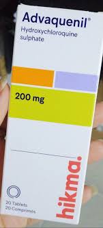
In the management of Systemic Lupus Erythematosus (SLE) complicated by cryoglobulinemia, a multidisciplinary approach is essential to address both systemic inflammatory control and localized musculoskeletal manifestations. Pharmacological intervention typically centers on disease-modifying therapies, including Advaquenil 200mg 200mg for baseline immunomodulation and Antiproliferative agents (e.g., Mycophenolate Mofetil, Azathioprine) / العوامل المضادة للتكاثر (مثل مايكوفينولات موفيتيل، أزاثيوبرين) Standard to manage refractory organ involvement and vasculitic complications. Beyond systemic therapy, clinicians must remain vigilant regarding the structural consequences of chronic inflammation, as detailed in Orthopaedic Management of Systemic Arthropathies: Reiter Syndrome, Gout, and Systemic Lupus Erythematosus. When joint degradation or hand deformities progress despite optimal medical management, surgical consultation is warranted, with specific considerations for Operative Management of Hand Arthritis: Osteoarthritis, Rheumatoid Arthritis, and SLE and differential diagnostic pathways for related inflammatory conditions such as those discussed in Surgical Management of Psoriatic Arthritis and Reiter Syndrome in the Hand.