Clinical Assessment & Protocol
Typical Presentation (HPI)
EN: Patient reports medial knee pain, stiffness after inactivity, and crepitus. AR: المريض يبلغ عن ألم في الجزء الأنسي من الركبة، تيبس بعد الخمول، وأصوات طقطقة.
General Examination
EN: Unremarkable or not routinely indicated. AR: طبيعي أو غير مطلوب روتينياً.
Treatment Protocol
EN: Strengthening of knee extensors, weight management, and activity modification. AR: تقوية باسطات الركبة، إدارة الوزن، وتعديل النشاط.
Patient Education
EN: Joint protection techniques and home exercise compliance. AR: تقنيات حماية المفاصل والالتزام بالتمارين المنزلية.
Systemic & Specialized Examinations
EN: S1, S2 present. No murmurs. AR: صوتا القلب الأول والثاني طبيعيان. لا توجد نفخات.
EN: Lungs clear to auscultation. AR: الرئتان صافيتان عند التسمع.
EN: Abdomen soft, non-tender. AR: البطن لين ولا يوجد ألم.
EN: Alert, oriented x3. No focal deficits. AR: المريض واعي ومدرك. لا يوجد عجز عصبي بؤري.
EN: Unremarkable or not routinely indicated. AR: طبيعي أو غير مطلوب روتينياً.
EN: Unremarkable or not routinely indicated. AR: طبيعي أو غير مطلوب روتينياً.
EN: Unremarkable or not routinely indicated. AR: طبيعي أو غير مطلوب روتينياً.
EN: Unremarkable or not routinely indicated. AR: طبيعي أو غير مطلوب روتينياً.
EN: Unremarkable or not routinely indicated. AR: طبيعي أو غير مطلوب روتينياً.
Orthopedic & Trauma Assessments
EN: Joint line tenderness, limited range of motion, and quadriceps atrophy. AR: إيلام عند خط المفصل، محدودية في مدى الحركة، وضمور في العضلة الرباعية.
EN: Unremarkable or not routinely indicated. AR: طبيعي أو غير مطلوب روتينياً.
Comprehensive Clinical Guide: Medial Compartment Osteoarthritis of the Knee
Osteoarthritis (OA) of the knee is a degenerative, chronic condition characterized by the progressive loss of articular cartilage, subchondral bone remodeling, and secondary inflammatory changes. While the knee joint comprises three primary compartments (medial, lateral, and patellofemoral), the medial compartment is the most frequently affected site due to the physiological distribution of weight-bearing forces. This guide provides a deep-dive into the clinical management, pathophysiology, and diagnostic standards for Medial Compartment Osteoarthritis (MCOA).
1. Clinical Definition and Overview
Medial Compartment Osteoarthritis (MCOA) is a mechanical and biochemical failure of the joint’s integrity, specifically involving the medial femoral condyle and the medial tibial plateau. Unlike generalized knee OA, MCOA is highly influenced by the "varus" alignment of the lower extremity, which places an asymmetric load on the medial aspect of the joint.
The Biomechanical Reality
In a healthy knee, forces are distributed relatively evenly. However, due to the natural anatomical alignment of the human leg, the medial compartment routinely bears approximately 60% to 80% of the total compressive load during the stance phase of gait. When this mechanical equilibrium is disrupted by injury, obesity, or genetic predisposition, the medial compartment undergoes rapid degradation.
2. Pathophysiology and Etiology
The transition from healthy hyaline cartilage to osteoarthritic degradation involves a complex cascade of molecular and mechanical events.
The Pathophysiological Cascade
- Chondrocyte Dysfunction: Initial insult leads to the activation of chondrocytes, which attempt to repair the matrix but instead produce pro-inflammatory cytokines (IL-1, TNF-alpha).
- Matrix Degradation: Increased production of matrix metalloproteinases (MMPs) leads to the breakdown of Type II collagen and proteoglycans.
- Subchondral Bone Remodeling: As cartilage thins, the underlying bone experiences increased stress, leading to sclerosis (hardening) and the formation of osteophytes (bone spurs) at the joint margins.
- Synovial Inflammation: Synovitis occurs as debris from cartilage breakdown triggers an immune response, leading to joint effusion and pain.
Etiological Factors
| Factor | Description |
|---|---|
| Varus Malalignment | Bow-legged deformity that shifts the mechanical axis medially. |
| Meniscal Deficiency | Prior medial meniscectomy or degenerative meniscal tears. |
| Obesity | Increased vertical load and systemic metabolic inflammation. |
| Genetics | Family history of early-onset cartilage degeneration. |
| Trauma | History of ACL or MCL injuries leading to instability. |
3. Clinical Staging and Grading: The Kellgren-Lawrence Scale
Standardized staging is critical for determining the therapeutic path. The Kellgren-Lawrence (KL) system remains the gold standard for radiographic classification.
| Grade | Description |
|---|---|
| 0 | No radiographic features of OA. |
| 1 | Doubtful joint space narrowing (JSN), possible osteophytic lipping. |
| 2 | Definite osteophytes, unimpaired joint space. |
| 3 | Moderate JSN, multiple osteophytes, mild sclerosis. |
| 4 | Large osteophytes, marked JSN, severe sclerosis, bone deformity. |
4. Standard Clinical Presentation
Patients typically present with a constellation of symptoms that correlate with both mechanical wear and inflammatory flares.
- Pain: Localized to the medial joint line. Often exacerbated by prolonged standing, stair climbing, and rising from a seated position.
- Stiffness: Morning stiffness lasting <30 minutes.
- Crepitus: Audible or palpable grinding during range of motion (ROM).
- Effusion: Periodic "swelling" of the knee, particularly after high-activity days.
- Mechanical Symptoms: Intermittent locking or catching (often indicating a concurrent degenerative meniscus tear).
- Deformity: Visible varus (bow-legged) thrust during gait.
5. Diagnostic Methodology
A systematic approach is required to differentiate MCOA from other pathologies.
Physical Examination
- Palpation: Tenderness specifically along the medial joint line.
- Range of Motion: Loss of full extension or flexion.
- Alignment Check: Evaluation of the mechanical axis (Varus vs. Valgus).
- Special Tests: McMurray’s test (for meniscal involvement) and the Varus Stress Test (to evaluate medial collateral ligament integrity).
Imaging Modalities
- Weight-Bearing Radiographs (Standing AP): Essential for visualizing JSN.
- Rosenberg View (45° Flexion): Superior for detecting early narrowing that standard views might miss.
- MRI: Rarely needed for diagnosis but useful if surgery is planned or if internal derangement (meniscus/ligament) is suspected.
6. Differential Diagnosis
It is imperative to exclude conditions that mimic MCOA:
* Rheumatoid Arthritis: Usually bilateral and systemic.
* Medial Meniscal Tear: Often acute or sub-acute onset, localized sharp pain.
* Pes Anserine Bursitis: Pain inferior to the joint line, usually responsive to local injection.
* Avascular Necrosis (AVN): Typically presents with sudden, severe, non-mechanical pain.
* Referred Pain: Hip pathology (e.g., hip OA) frequently presents as knee pain.
7. Risks, Contraindications, and Long-Term Prognosis
Risks of Untreated MCOA
- Gait Alteration: Compensatory changes in the hip and ankle.
- Muscle Atrophy: Quadriceps inhibition leads to decreased stability.
- Permanent Deformity: Progression to severe varus deformity requiring complex arthroplasty.
Contraindications for Conservative Management
- Presence of severe neurological compromise.
- Infection (Septic Arthritis).
- Failure of conservative measures (typically 6 months of dedicated therapy) in the presence of KL Grade 4 disease.
Prognosis
While OA is a progressive, irreversible disease, it is highly manageable. Early intervention—specifically weight loss and physical therapy—can delay the need for surgical intervention by a decade or more.
8. Frequently Asked Questions (FAQ)
1. Is surgery the only option for Medial Compartment OA?
No. Surgery is typically reserved for cases where conservative management (PT, weight loss, bracing) fails to provide an acceptable quality of life.
2. Can I reverse cartilage loss?
Currently, there is no clinically proven method to regenerate hyaline cartilage in an adult knee. Treatment focuses on symptom management and slowing progression.
3. Does wearing a brace help?
Yes. An "unloader brace" is specifically designed to shift weight away from the medial compartment, providing significant pain relief for patients with varus alignment.
4. What is the role of injections?
Hyaluronic acid (viscosupplementation) and corticosteroids provide temporary relief. Corticosteroids are best for acute flares, while HA may provide longer-term lubrication for mild-to-moderate cases.
5. Is exercise harmful to my knee?
On the contrary, low-impact exercise (swimming, cycling, elliptical) is vital. It maintains joint lubrication, strengthens supporting musculature, and aids in weight management.
6. Why does my knee "click"?
Crepitus is often caused by cartilage surfaces rubbing against each other or by small bone spurs. As long as it is not accompanied by locking or severe pain, it is often a benign symptom of aging joints.
7. How much weight loss is required to see a difference?
Losing even 5-10% of body weight can reduce the compressive force on the knee by several times that amount during walking, significantly lowering pain levels.
8. What is a high tibial osteotomy (HTO)?
HTO is a surgical procedure for younger, active patients that realigns the leg to shift weight from the damaged medial compartment to the healthy lateral compartment.
9. Can MCOA lead to hip problems?
Yes. Altered gait patterns due to knee pain can lead to secondary biomechanical stress in the hip and lower back.
10. How often should I see an orthopedic specialist?
Patients with known MCOA should have annual follow-ups to monitor progression, unless symptoms worsen significantly (e.g., sudden swelling or loss of motion).
9. Clinical Management Strategies
Conservative Protocols
- Physical Therapy: Focus on quadriceps strengthening (specifically the VMO) and hamstring flexibility.
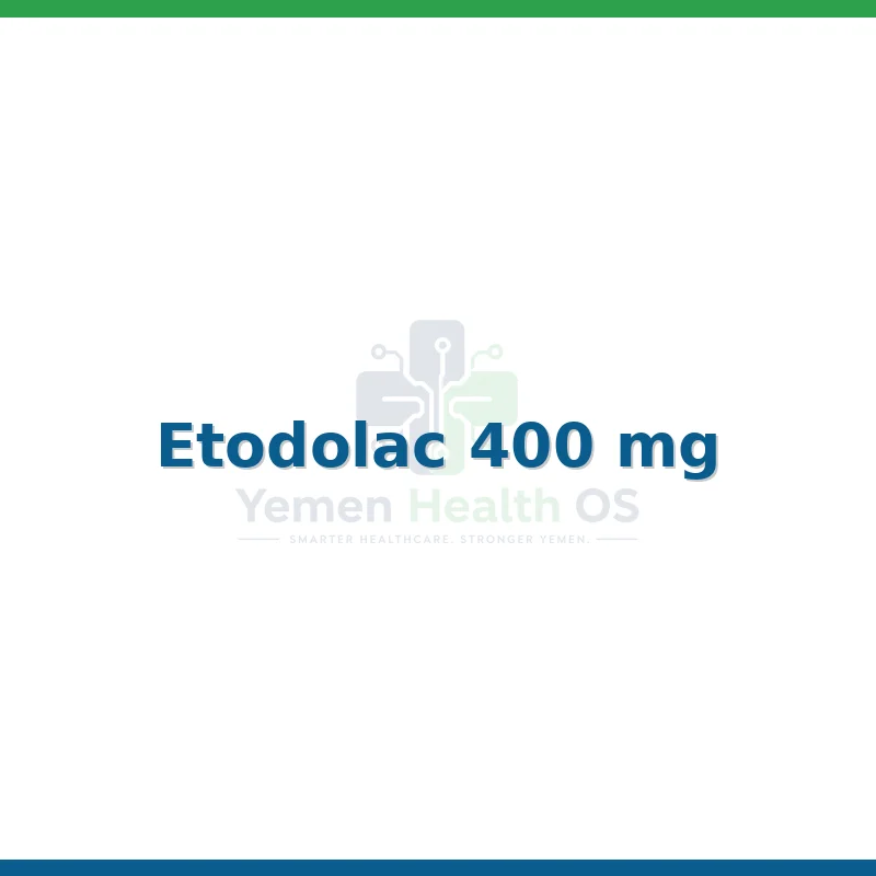
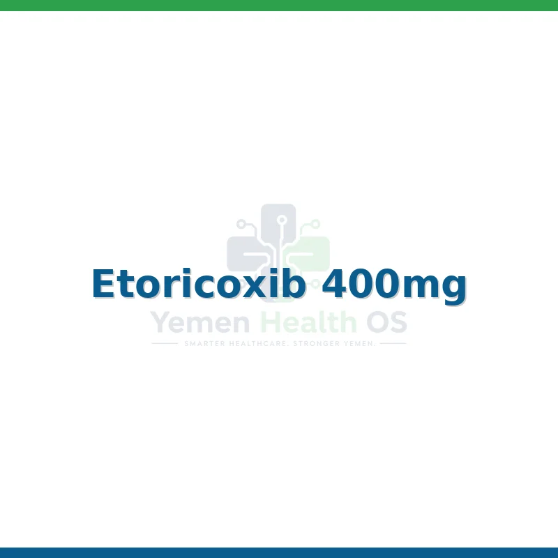
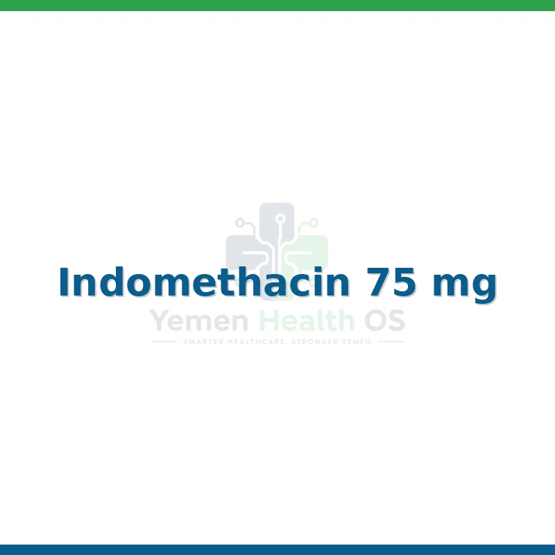
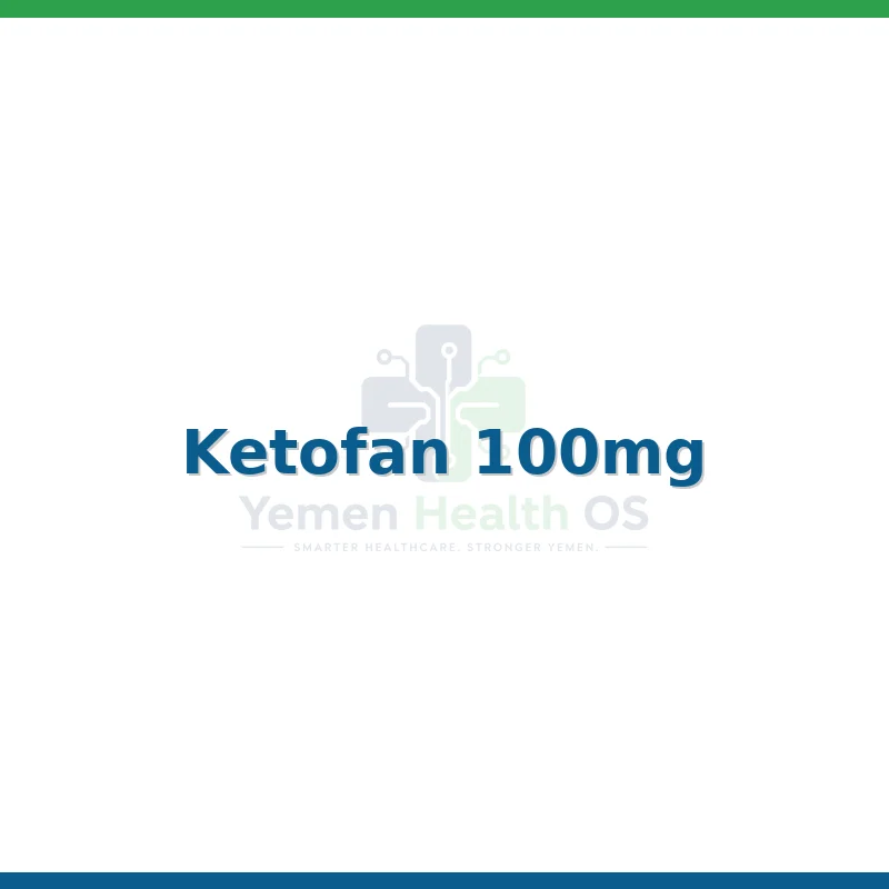
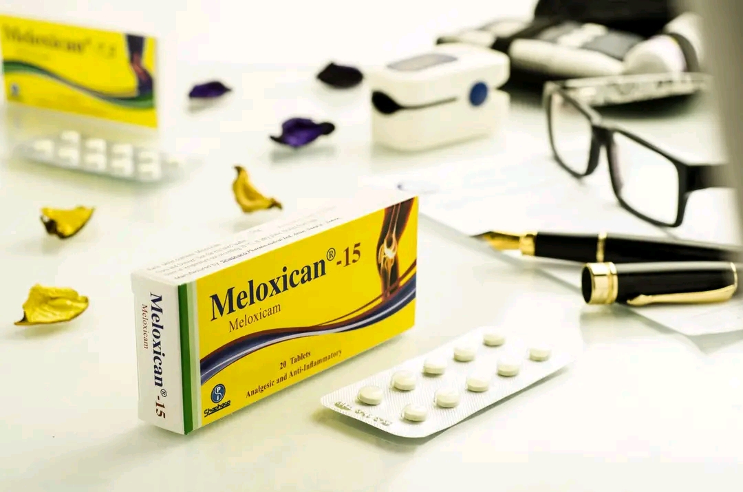
- Pharmacology: NSAIDs (topical or oral) are first-line. Acetaminophen is an alternative for those with cardiovascular or GI contraindications.
- Orthotics: Lateral wedge insoles can help reduce the varus moment during the gait cycle.
Surgical Interventions
- Arthroscopic Debridement: Generally discouraged for pure OA, but useful if a mechanical meniscal flap is present.
- Unicompartmental Knee Arthroplasty (UKA): A minimally invasive option for patients with isolated medial disease.
- Total Knee Arthroplasty (TKA): The gold standard for end-stage, multi-compartment disease, offering high long-term success rates.
Conclusion
Medial Compartment Osteoarthritis is a multifaceted condition that requires a patient-centered approach. By understanding the mechanical drivers—specifically the varus alignment—clinicians can better tailor interventions. Early recognition, combined with a robust conservative strategy involving physiotherapy and lifestyle modification, remains the cornerstone of effective management, ensuring patients maintain mobility and quality of life for as long as possible before considering surgical intervention.
Treatment & Management Options
Recommended Medications