Clinical Assessment & Protocol
Typical Presentation (HPI)
EN: Patient presents for evaluation of persistent pain and functional limitation at the site of a previous left tibial shaft fracture. Fracture occurred [Date], treated initially with [ORIF/IMN/Cast]. Patient reports localized tenderness, inability to bear full weight without discomfort, and lack of clinical or radiographic progression toward union over the past [Number] months. No systemic symptoms of infection (fever, chills, night sweats) reported. AR: يراجع المريض لتقييم الألم المستمر ومحدودية الوظيفة في موقع كسر سابق في ساق الظنبوب الأيسر. حدث الكسر بتاريخ [التاريخ]، وتم علاجه مبدئياً بـ [تثبيت جراحي داخلي/مسمار نخاعي/جبيرة]. يشكو المريض من ألم موضعي، وعدم القدرة على تحمل الوزن الكامل دون انزعاج، وغياب أي تقدم سريري أو شعاعي نحو الالتئام خلال الأشهر [العدد] الماضية. لا توجد أعراض جهازية للعدوى (حمى، قشعريرة، تعرق ليلي).
General Examination
EN: Left lower extremity examination reveals localized tenderness at the fracture site. No erythema, warmth, or sinus tract formation noted. Range of motion at the knee and ankle is [Full/Restricted]. Neurovascular status is intact with palpable dorsalis pedis and posterior tibial pulses; capillary refill < 2 seconds. Radiographic assessment demonstrates persistent fracture gap, lack of bridging callus, and sclerotic bone ends consistent with nonunion. AR: فحص الطرف السفلي الأيسر يكشف عن ألم موضعي عند موقع الكسر. لا توجد علامات احمرار أو حرارة أو تشكل مسارات ناسويه. مدى الحركة في الركبة والكاحل [كامل/محدود]. الحالة العصبية الوعائية سليمة مع وجود نبضات الشريان ظهر القدم والشريان الظنبوبي الخلفي؛ زمن إعادة التعبئة الشعرية أقل من ثانيتين. التقييم الشعاعي يظهر فجوة مستمرة في الكسر، وغياب الدشبذ العظمي الجسري، وتصلب نهايات العظام بما يتوافق مع عدم الالتئام.
Treatment Protocol
EN: Plan: 1. Obtain inflammatory markers (ESR, CRP) to rule out occult infection. 2. Discuss surgical revision options, including exchange nailing, bone grafting (autograft/allograft), or compression plating. 3. Consider adjunctive therapies such as low-intensity pulsed ultrasound (LIPUS) or bone stimulators. 4. Maintain protected weight-bearing as tolerated with assistive devices. 5. Follow-up in [Number] weeks for repeat imaging. AR: الخطة: 1. طلب تحاليل المؤشرات الالتهابية (ESR, CRP) لاستبعاد وجود عدوى خفية. 2. مناقشة خيارات المراجعة الجراحية، بما في ذلك استبدال المسمار النخاعي، أو تطعيم العظام (ذاتي/خارجي)، أو التثبيت بالصفائح الضاغطة. 3. النظر في العلاجات المساعدة مثل الموجات فوق الصوتية النبضية منخفضة الكثافة (LIPUS) أو محفزات نمو العظام. 4. الحفاظ على تحمل الوزن المحمي حسب القدرة باستخدام أدوات مساعدة. 5. المتابعة بعد [العدد] أسابيع لإعادة التصوير الشعاعي.
Patient Education
EN: Nonunion means the bone has failed to heal within the expected timeframe. This may be due to biological factors, mechanical instability, or blood supply issues. It is critical to avoid smoking, as nicotine significantly impairs bone healing. Please adhere to your weight-bearing restrictions to prevent further hardware failure or displacement. Report any sudden increase in pain, drainage, or fever immediately. AR: عدم الالتئام يعني أن العظم لم يلتئم ضمن الإطار الزمني المتوقع. قد يعود ذلك لعوامل بيولوجية، أو عدم استقرار ميكانيكي، أو مشاكل في التروية الدموية. من الضروري جداً تجنب التدخين، حيث أن النيكوتين يعيق التئام العظام بشكل كبير. يرجى الالتزام بقيود تحمل الوزن الموصى بها لمنع فشل الأدوات الجراحية أو إزاحة الكسر. يرجى إبلاغنا فوراً في حال حدوث زيادة مفاجئة في الألم، أو وجود إفرازات، أو ارتفاع في درجة الحرارة.
Systemic & Specialized Examinations
EN: Distal neurovascular assessment is critical: INTACT. No signs of acute compartment syndrome (Pain on passive toe stretch is negative). AR: التقييم العصبي الوعائي الطرفي سليم. لا توجد علامات لمتلازمة الحيز الحادة (ألم عند الشد السلبي للأصابع سلبي).
Orthopedic & Trauma Assessments
EN: High-impact direct blow or severe torsional forces. AR: ضربة مباشرة قوية أو قوى التواء شديدة.
EN: Non-ambulatory. Arrived via EMS/stretcher. AR: غير قادر على المشي. وصل عبر الإسعاف/نقالة.
EN: Marked soft tissue swelling and ecchymosis. The limb appears shortened and externally rotated (if femur/hip) or grossly angulated (if tibia). AR: تورم ملحوظ وكدمات. يبدو الطرف أقصر ومستدار للخارج (إذا كان الفخذ/الورك) أو مقوس بشكل كبير (إذا كان الظنبوب).
EN: N/A in acute fracture. AR: لا ينطبق.
EN: Distal toes move symmetrically. EHL/FHL intact. AR: أصابع القدم تتحرك بتمائل. باسطة/قابضة الإبهام سليمة.
EN: Sensation intact to light touch in all distal dermatomes (Peroneal and Tibial nerves). AR: الإحساس سليم للمس الخفيف في جميع المناطق الطرفية (العصب الشظوي والظنبوبي).
EN: Deferred. AR: مؤجل.
EN: DP and PT pulses are strong, bounding 2+. Capillary refill < 2 seconds. AR: النبضات الطرفية قوية 2+. عودة امتلاء الشعيرات سريعة.
Clinical Guide: Nonunion of Tibial Shaft Fracture, Left Leg
1. Comprehensive Introduction & Overview
Nonunion of a tibial shaft fracture is a significant orthopedic complication characterized by the cessation of all reparative processes in a fracture site without bony union. The tibia, being the most commonly fractured long bone in the human body, is uniquely susceptible to nonunion due to its precarious vascular supply, particularly in the distal third. A diagnosis of "Nonunion of Tibial Shaft Fracture, Left Leg" implies that the fracture has failed to heal within the expected timeframe—typically defined as six to nine months post-injury—and that the biological and mechanical environment is insufficient to achieve union without clinical intervention.
This guide serves as a technical resource for clinical specialists, providing an exhaustive overview of the pathophysiology, diagnostic criteria, and management frameworks required to address this challenging clinical entity.
2. Deep-Dive: Technical Specifications and Mechanisms
Etiology and Pathophysiological Drivers
The tibia is a subcutaneous bone with a high-energy mechanism of injury (often motor vehicle accidents or falls) frequently resulting in soft tissue compromise. The failure of union is categorized into two primary biological states:
- Hypertrophic Nonunion (The "Elephant Foot"): Characterized by abundant callus formation. The biological potential for healing is present, but mechanical stability is insufficient.
- Atrophic Nonunion: Characterized by a lack of callus formation. This is a biological failure, often due to poor vascularity, systemic factors, or soft tissue interposition.
The Role of Vascularity
The blood supply to the tibial shaft is primarily derived from the nutrient artery entering the posterior cortex. High-energy fractures often disrupt the endosteal blood supply, leaving the bone dependent on the periosteal blood supply. If the periosteum is stripped during the initial injury or surgical exposure, the bone becomes avascular, leading to necrosis and subsequent atrophic nonunion.
Mechanical Instability
Micromotion at the fracture site is necessary for secondary bone healing (callus formation). However, excessive motion (macromotion) disrupts the formation of the bridging callus and promotes the development of fibrous tissue rather than bone.
| Factor | Impact on Healing |
|---|---|
| Smoking/Nicotine | Vasoconstriction, reduces oxygen tension at fracture site. |
| Diabetes Mellitus | Impairs collagen synthesis and osteoblast function. |
| Infection | Induces inflammatory cytokines that inhibit osteogenesis. |
| Malalignment | Creates shear forces that prevent bridging callus. |
3. Clinical Staging and Grading
Orthopedic specialists typically utilize the Weber and Cech Classification to determine the therapeutic approach:
Hypertrophic (Biological Potential High)
- Type: Elephant Foot, Horse Hoof, Oligotrophic.
- Management: Improving mechanical stability (e.g., dynamization or exchange nailing).
Atrophic (Biological Potential Low)
- Type: Torsion Wedge, Comminuted, Defect, Atrophic.
- Management: Biological stimulation (bone grafting, BMPs) combined with rigid fixation.
4. Standard Presentation and Differential Diagnosis
Clinical Presentation
Patients presenting with a suspected nonunion of the left tibial shaft will typically report:
* Persistent Pain: Localized pain at the fracture site, often exacerbated by weight-bearing.
* Deformity: Visible angulation or rotational malalignment.
* Instability: A palpable "click" or movement at the fracture site during clinical examination.
* Soft Tissue Status: Possible history of draining sinuses (if infection is present).
Differential Diagnosis
- Delayed Union: A fracture that is healing but at a slower rate than expected (not yet a nonunion).
- Osteomyelitis: A chronic bone infection mimicking nonunion pain.
- Complex Regional Pain Syndrome (CRPS): Can mimic the pain profile of a nonunion.
- Hardware Failure: Fatigue or breakage of the intramedullary nail or plates.
5. Key Diagnostic Tests
To confirm nonunion, a multi-modal diagnostic approach is required:
- Radiographic Imaging (AP/Lateral): Assessment of callus, gap size, and hardware integrity.
- CT Scan: The gold standard for assessing bridging bone. It identifies the presence of cortices across the fracture gap.
- Laboratory Markers: ESR (Erythrocyte Sedimentation Rate) and CRP (C-Reactive Protein) are essential to rule out occult infection.
- Stress Views: Fluoroscopic assessment under stress to detect abnormal motion at the fracture site.
- Bone Scintigraphy/PET Scan: Occasionally used to differentiate between metabolic activity (hypertrophic) and biological inactivity (atrophic).
6. Clinical Indications and Usage: Management Strategies
Surgical Intervention
- Exchange Nailing: Removing the current intramedullary nail, reaming the canal to increase the diameter, and inserting a larger nail to increase stability and provide autologous bone graft (via reamer-irrigator-aspirator).
- Plate Fixation: Used when intramedullary nailing fails or when the fracture is near the metaphysis.
- Bone Grafting: Autograft (iliac crest) remains the gold standard, though synthetic bone graft substitutes (BMP-2) are increasingly utilized in atrophic cases.
Non-Surgical Adjuncts
- Pulsed Electromagnetic Field (PEMF) Therapy: Utilized in select cases to stimulate osteoblastic activity.
- Low-Intensity Pulsed Ultrasound (LIPUS): May be indicated for delayed unions or post-surgical support.
7. Risks, Side Effects, and Contraindications
Risks of Surgical Revision
- Deep Infection: A catastrophic complication requiring hardware removal and potential antibiotic bead therapy.
- Neurovascular Injury: Risk to the peroneal nerve during hardware placement or revision.
- Compartment Syndrome: High-risk in revision surgery due to existing scarring and altered anatomy.
Contraindications for Revision
- Active Infection: Must be cleared prior to internal fixation.
- Severe Systemic Illness: Patients who are not medically stable for prolonged anesthesia.
- Non-compliant Patients: If the patient cannot adhere to weight-bearing restrictions.
8. Long-Term Prognosis
The prognosis for nonunion of the tibial shaft is generally favorable with appropriate surgical intervention, though it often requires a prolonged recovery period. Factors influencing success include:
* Resolution of Infection: If present, the prognosis is guarded until the infection is eradicated.
* Biological Viability: Patients who cease nicotine use and optimize glycemic control have significantly higher success rates.
* Mechanical Environment: Achieving rigid stability is the single most important factor for union.
9. Massive FAQ Section
Q1: How long must a fracture remain unhealed before it is called a "nonunion"?
A: Generally, if a fracture shows no signs of healing on radiographs after 6 to 9 months, it is clinically classified as a nonunion.
Q2: Does smoking really affect the healing of my left tibia?
A: Yes. Nicotine is a potent vasoconstrictor that reduces the delivery of oxygen and nutrients to the fracture site, significantly increasing the risk of atrophic nonunion.
Q3: What is the difference between delayed union and nonunion?
A: Delayed union is a fracture that is taking longer than average to heal but is still showing progressive signs of callus formation. Nonunion is a state where the healing process has completely stalled.
Q4: Why is the left tibia harder to heal than other bones?
A: The tibia has a thin soft-tissue envelope and a precarious blood supply. High-energy trauma often damages these vessels, leading to poor healing.
Q5: Will I need a bone graft?
A: If the nonunion is atrophic (lacking biological activity), a bone graft is often necessary to provide the "scaffold" and biological signal for new bone growth.
Q6: Can I walk on a nonunion?
A: Walking on a nonunion without physician clearance can lead to hardware failure, further deformity, or propagation of the nonunion. Always follow the weight-bearing protocols set by your surgeon.
Q7: What are the signs of an infected nonunion?
A: Persistent pain, warmth, redness, and potentially a draining sinus tract (a sore that won't heal) are indicators of infection.
Q8: Is PEMF therapy effective?
A: PEMF is a non-invasive option primarily used for hypertrophic nonunions where mechanical stability has been achieved but biological stimulation is required.
Q9: What is "Exchange Nailing"?
A: It is a procedure where an existing intramedullary nail is removed, the bone canal is widened (reamed), and a larger nail is inserted to improve stability.
Q10: How do I know if my nonunion is "atrophic" or "hypertrophic"?
A: Your surgeon will determine this via radiographs. Hypertrophic nonunions show a "callus cloud" (too much bone, not enough stability), whereas atrophic nonunions show a "gap" or "rounded edges" (no callus, no biological activity).
10. Conclusion
The management of a Nonunion of the Tibial Shaft Fracture, Left Leg, represents a complex intersection of biology and mechanics. Success relies on accurate classification, the eradication of infection, the restoration of mechanical stability, and the optimization of the patient’s biological environment. By adhering to standardized evidence-based protocols, orthopedic specialists can significantly improve clinical outcomes and restore function to the patient's lower limb.
Disclaimer: This guide is for educational and clinical reference purposes only and does not supersede professional medical judgment. Always consult with a board-certified orthopedic surgeon regarding specific patient cases.
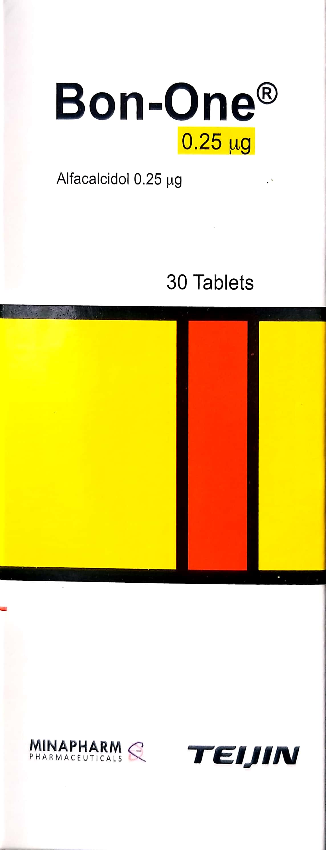
Related Clinical Integration
The management of a Nonunion of Tibial Shaft Fracture, Left Leg requires a multidisciplinary approach that integrates advanced surgical intervention, pharmacological support, and patient-centered rehabilitation. Clinicians often utilize a Battery Powered Orthopedic Drill/Saw System to facilitate precise hardware revision or debridement, while procedures such as Alveolar Bone Grafting—or analogous autologous bone grafting techniques—are essential for stimulating osteogenesis in atrophic nonunions. To optimize the systemic environment for bone healing, practitioners may prescribe Calcium Gluconate / غلوكونات الكالسيوم 10ml and Bon-one / بون-ون 0.25mcg to address metabolic deficiencies, while ensuring patient mobility through the use of Axillary (Underarm) Crutches / عكازات إبطية (أدوات ومساعدات الحركة (عكازات/كراسي)). For a deeper understanding of the clinical decision-making process, providers should consult resources on Nonunion Fractures: Orthopedic Epidemiology, Biomechanics, & Classification, Advanced Management of Fracture Nonunions: Biomechanics, Bone Grafting, and Surgical Reconstruction, and Mastering the Surgical Management of Lower Extremity Nonunions: Tibia and Foot. Furthermore, specialized techniques such as Ilizarov External Fixation for Tibial Shaft Fractures: Surgical Guide and comprehensive