Clinical Assessment & Protocol
Typical Presentation (HPI)
EN: High fever, dry cough, and gastrointestinal symptoms (diarrhea) in a hotel guest. AR: حمى عالية، وسعال جاف، وأعراض هضمية (إسهال) لدى نزيل في فندق.
General Examination
EN: Relative bradycardia, pulmonary consolidation, and confusion. AR: بطء نبض نسبي، وتصلد رئوي، وارتباك.
Treatment Protocol
EN: Fluoroquinolones (levofloxacin) or macrolides (azithromycin). AR: فلوروكوينولونات (ليفوفلوكساسين) أو ماكروليدات (أزيثروميسين).
Patient Education
EN: Report potential outbreaks related to building water systems to local health authorities. AR: إبلاغ السلطات الصحية المحلية عن التفشيات المحتملة المرتبطة بأنظمة مياه المباني.
Systemic & Specialized Examinations
EN: S1, S2 present. No murmurs. AR: صوتا القلب الأول والثاني طبيعيان. لا توجد نفخات.
EN: Lungs clear to auscultation. AR: الرئتان صافيتان عند التسمع.
EN: Abdomen soft, non-tender. AR: البطن لين ولا يوجد ألم.
EN: Alert, oriented x3. No focal deficits. AR: المريض واعي ومدرك. لا يوجد عجز عصبي بؤري.
EN: Unremarkable or not routinely indicated. AR: طبيعي أو غير مطلوب روتينياً.
EN: Unremarkable or not routinely indicated. AR: طبيعي أو غير مطلوب روتينياً.
EN: Unremarkable or not routinely indicated. AR: طبيعي أو غير مطلوب روتينياً.
EN: Unremarkable or not routinely indicated. AR: طبيعي أو غير مطلوب روتينياً.
EN: Unremarkable or not routinely indicated. AR: طبيعي أو غير مطلوب روتينياً.
Orthopedic & Trauma Assessments
EN: Unremarkable or not routinely indicated. AR: طبيعي أو غير مطلوب روتينياً.
EN: Unremarkable or not routinely indicated. AR: طبيعي أو غير مطلوب روتينياً.
Comprehensive Clinical Guide: Legionnaires’ Disease (Legionella pneumophila)
1. Introduction and Clinical Overview
Legionnaires’ disease is a severe, systemic form of atypical pneumonia caused by the gram-negative, aerobic bacterium Legionella pneumophila. First identified following a 1976 outbreak at an American Legion convention in Philadelphia, this pathogen remains a significant public health concern. Unlike community-acquired pneumonia (CAP) caused by common pathogens like Streptococcus pneumoniae, Legionellosis presents with distinct multisystem involvement, often complicating clinical management.
The disease manifests in two primary clinical forms:
* Legionnaires’ Disease: The severe, pneumonia-inducing form characterized by high fever, pulmonary consolidation, and potential respiratory failure.
* Pontiac Fever: A self-limiting, non-pneumonic, flu-like illness characterized by fever, myalgia, and malaise, typically resolving within 2–5 days without antibiotic intervention.
This guide focuses specifically on the severe pneumonic form, which requires aggressive pharmacological intervention and high-acuity monitoring.
2. Etiology and Pathophysiology
Etiology
Legionella species are ubiquitous in freshwater environments, including lakes and streams. However, the primary clinical concern arises when these organisms colonize man-made water systems, such as:
* Cooling towers and evaporative condensers.
* Hot tubs and whirlpool spas.
* Complex hospital plumbing systems (showers, faucets).
* Decorative fountains and misting machines.
Transmission occurs via the inhalation of aerosolized water droplets containing the bacteria. Notably, there is no evidence of person-to-person transmission.
Pathophysiology
The virulence of L. pneumophila is rooted in its ability to survive and replicate within human alveolar macrophages. The mechanism follows a distinct molecular cascade:
1. Inhalation: Aerosolized bacteria reach the lower respiratory tract.
2. Phagocytosis: The bacteria are engulfed by alveolar macrophages.
3. Intracellular Inhibition: The bacteria secrete proteins via the Dot/Icm (Defect in organelle trafficking/Intracellular multiplication) type IV secretion system. This prevents the phagosome-lysosome fusion, creating a "Legionella-containing vacuole" (LCV).
4. Replication: The bacterium replicates within the LCV, eventually causing cell lysis and the release of progeny into the lung parenchyma.
5. Inflammatory Response: The host immune response triggers a robust influx of neutrophils and cytokines (IL-1, IL-6, TNF-alpha), leading to the characteristic alveolar consolidation seen in radiographic imaging.
3. Clinical Staging and Standard Presentation
Legionnaires’ disease often presents with a deceptive clinical course, starting with mild symptoms and rapidly progressing to systemic distress.
| Stage | Manifestations |
|---|---|
| Prodrome (Days 1–2) | Malaise, myalgia, low-grade fever, mild headache. |
| Acute Phase (Days 3–6) | High fever (>39°C), productive/non-productive cough, rigors, dyspnea. |
| Systemic Phase | Gastrointestinal distress (diarrhea, nausea), confusion/altered mental status, relative bradycardia. |
Key Clinical Indicators
- Respiratory: Pleuritic chest pain, hemoptysis, hypoxemia.
- Neurological: Confusion, lethargy, or ataxia (often more pronounced than in other bacterial pneumonias).
- Gastrointestinal: Watery diarrhea occurs in approximately 25–50% of patients.
- Laboratory findings: Hyponatremia (often <130 mEq/L), elevated hepatic transaminases, and elevated CRP/Procalcitonin.
4. Differential Diagnosis
Because the clinical presentation mimics other infectious and inflammatory conditions, clinicians must maintain a high index of suspicion:
- Streptococcus pneumoniae: Typically presents with more acute lobar consolidation and "rust-colored" sputum.
- Mycoplasma pneumoniae: Often affects younger populations with a more protracted, "walking pneumonia" course.
- Viral Pneumonia (Influenza/COVID-19): Often bilateral, diffuse ground-glass opacities; lacks the specific gastrointestinal and hyponatremic profile of Legionellosis.
- Psittacosis/Q Fever: Zoonotic exposures should be considered if the patient has contact with animals.
- Non-infectious causes: Pulmonary embolism, congestive heart failure, or drug-induced pneumonitis.
5. Diagnostic Testing Protocols
The gold standard for diagnosis requires a multi-modal approach, as traditional sputum cultures are notoriously insensitive.
- Urinary Antigen Test (UAT): The most commonly used diagnostic tool. It detects L. pneumophila serogroup 1 antigen. It is rapid, highly specific, and remains positive for days to weeks after antibiotic therapy begins.
- Culture (Lower Respiratory Secretions): The definitive diagnostic method. Requires Buffered Charcoal Yeast Extract (BCYE) agar. It is the only method that can identify non-serogroup 1 strains.
- Nucleic Acid Amplification Tests (NAAT/PCR): Highly sensitive and specific. Preferred for rapid, broad-spectrum identification of Legionella species.
- Imaging: Chest X-rays typically show unilateral patchy infiltrates that may progress to consolidation. CT scans are superior for identifying early cavitation or pleural effusions.
6. Pharmacological Management and Risks
Standard Treatment
Legionella is an intracellular pathogen; therefore, antibiotics must demonstrate high intracellular penetration.
* First-line: Fluoroquinolones (Levofloxacin or Moxifloxacin) for 7–14 days.
* Alternative: Macrolides (Azithromycin) are often used, particularly in patients with contraindications to fluoroquinolones.
* Severe Cases: Combination therapy (e.g., a fluoroquinolone plus rifampin) may be considered in immunocompromised patients or those with severe respiratory failure.
Contraindications and Side Effects
- Fluoroquinolones: Risk of tendonitis, tendon rupture, peripheral neuropathy, and QTc prolongation. Monitor cardiac telemetry if the patient is on other QTc-prolonging agents.
- Macrolides: Gastrointestinal distress and potential for cardiac arrhythmia (torsades de pointes).
7. Long-Term Prognosis and Recovery
While mortality in treated, healthy individuals is low (approx. 5–10%), it can rise significantly in elderly patients or those with comorbid COPD/diabetes.
* Convalescence: Fatigue, lethargy, and neurocognitive deficits may persist for weeks or months after the infection has cleared.
* Pulmonary Function: Long-term pulmonary fibrosis is rare but possible in patients who required prolonged mechanical ventilation.
8. Frequently Asked Questions (FAQ)
1. Is Legionnaires’ disease contagious between people?
No. It is not transmitted from person to person. You acquire it by breathing in mist containing the bacteria.
2. How long does the incubation period last?
The incubation period is typically 2 to 10 days, though it can occasionally extend to 14 days.
3. Why does this disease cause hyponatremia?
The exact mechanism is debated, but it is likely linked to the Syndrome of Inappropriate Antidiuretic Hormone (SIADH) secretion associated with the severe systemic inflammatory response.
4. Can I get Legionnaires’ from drinking water?
No. The bacteria must be aspirated into the lungs. Drinking water is generally safe unless it is aspirated (choked on).
5. Is there a vaccine for Legionnaires’ disease?
Currently, there is no commercially available vaccine for Legionella.
6. Who is at the highest risk?
Individuals over 50, current or former smokers, those with chronic lung disease, and immunocompromised patients (e.g., transplant recipients).
7. How do hospitals prevent outbreaks?
Hospitals implement strict water management programs (WMPs) involving temperature control, chlorine residual monitoring, and UV disinfection of water lines.
8. Is the Urinary Antigen Test enough for diagnosis?
It is excellent for identifying Serogroup 1 (the most common cause of outbreaks), but it will miss other serogroups. If clinical suspicion is high and the test is negative, further testing (PCR/culture) is required.
9. Can I return to work once I feel better?
Patients can usually return to work once their fever has subsided and their respiratory symptoms are improving, generally after 48–72 hours of appropriate antibiotic therapy, provided they are no longer clinically unstable.
10. What is the difference between Pontiac Fever and Legionnaires’?
Pontiac Fever is a milder, self-limiting form of the disease that does not involve lung infection (pneumonia) and does not require antibiotics.
9. Clinical Summary Table: Management Checklist
| Action Item | Clinical Rationale |
|---|---|
| Pulse Oximetry | Monitor for silent hypoxemia. |
| Electrolyte Panel | Track hyponatremia; address if symptomatic. |
| Liver Function Tests | Assess for transaminitis common in systemic infection. |
| Respiratory Isolation | Not required (no person-to-person spread). |
| Empiric Coverage | Ensure antibiotic covers intracellular activity. |
| Hydration | Essential for managing systemic toxicity and maintaining renal perfusion. |
Disclaimer: This guide is intended for educational purposes for healthcare professionals and students. It does not replace institutional clinical protocols or direct physician judgment. In the event of a suspected outbreak, immediate notification of local public health authorities is mandatory.
Related Clinical Integration
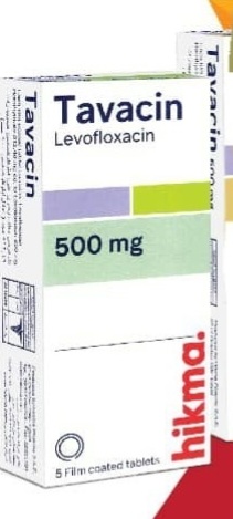
In the clinical management of Legionnaires' disease, diagnostic confirmation and targeted antimicrobial therapy are paramount for patient outcomes. Initial diagnostic workup typically necessitates Blood Cultures / مزارع الدم (خدمات رعاية عامة) and Urine culture and sensitivity testing / مزرعة البول واختبار الحساسية (خدمات رعاية عامة) to rule out co-infections and isolate the causative pathogen. Once the diagnosis is established, fluoroquinolones are considered the gold standard for treatment; clinicians may prescribe Lefoxin (Levofloxacin) / ليفوكسين (ليفوفلوكساسين) 500mg, Tavacin (Levofloxacin) / تافاسين (ليفوفلوكساسين) 500mg, or generic Levofloxacin / ليفوفلوكساسين 500mg to effectively penetrate intracellular reservoirs of Legionella pneumophila and ensure rapid clinical resolution.