Clinical Assessment & Protocol
Typical Presentation (HPI)
EN: Child presenting with bowed legs and delayed motor milestones. AR: طفل يعاني من تقوس الساقين وتأخر في المعالم الحركية.
General Examination
EN: Bowing of lower extremities, short stature, and dental abnormalities. AR: تقوس الأطراف السفلية، قصر القامة، وتشوهات في الأسنان.
Treatment Protocol
EN: Oral phosphate supplementation and active vitamin D analogues. AR: مكملات الفوسفات عن طريق الفم ونظائر فيتامين د النشطة.
Patient Education
EN: Monitor serum phosphate and renal ultrasound regularly. AR: مراقبة مستوى فوسفات الدم والأشعة التلفزيونية للكلية بانتظام.
Systemic & Specialized Examinations
EN: S1, S2 present. No murmurs. AR: صوتا القلب الأول والثاني طبيعيان. لا توجد نفخات.
EN: Lungs clear to auscultation. AR: الرئتان صافيتان عند التسمع.
EN: Abdomen soft, non-tender. AR: البطن لين ولا يوجد ألم.
EN: Alert, oriented x3. No focal deficits. AR: المريض واعي ومدرك. لا يوجد عجز عصبي بؤري.
EN: Unremarkable or not routinely indicated. AR: طبيعي أو غير مطلوب روتينياً.
EN: Unremarkable or not routinely indicated. AR: طبيعي أو غير مطلوب روتينياً.
EN: Unremarkable or not routinely indicated. AR: طبيعي أو غير مطلوب روتينياً.
EN: Unremarkable or not routinely indicated. AR: طبيعي أو غير مطلوب روتينياً.
EN: Unremarkable or not routinely indicated. AR: طبيعي أو غير مطلوب روتينياً.
Orthopedic & Trauma Assessments
EN: Unremarkable or not routinely indicated. AR: طبيعي أو غير مطلوب روتينياً.
EN: Unremarkable or not routinely indicated. AR: طبيعي أو غير مطلوب روتينياً.
Clinical Comprehensive Guide: Hypophosphatemic Rickets
1. Comprehensive Introduction & Overview
Hypophosphatemic Rickets (HR) represents a spectrum of rare metabolic bone disorders characterized by a chronic deficit in serum phosphate, leading to impaired mineralization of the growth plate cartilage and osteoid. Unlike nutritional rickets, which is primarily driven by Vitamin D deficiency, Hypophosphatemic Rickets is rooted in renal phosphate wasting.
The most common form is X-linked Hypophosphatemia (XLH), caused by mutations in the PHEX gene. However, the condition encompasses various genetic and acquired forms, all sharing the core pathophysiological hallmark of elevated Fibroblast Growth Factor 23 (FGF23) levels. If left untreated, the condition manifests as profound skeletal deformities, stunted growth, and lifelong musculoskeletal morbidity.
2. Technical Specifications & Pathophysiology
The FGF23 Axis
The crux of Hypophosphatemic Rickets lies in the dysregulation of phosphate homeostasis, mediated primarily by Fibroblast Growth Factor 23 (FGF23).
- Physiological Role: FGF23 is a hormone secreted by osteocytes that regulates phosphate excretion in the proximal convoluted tubule of the kidney.
- Pathological Mechanism: In HR, FGF23 levels are inappropriately elevated, leading to:
- Downregulation of NPT2a and NPT2c: These are sodium-phosphate cotransporters. Their reduction causes massive phosphate wasting (phosphaturia).
- Inhibition of 1-alpha-hydroxylase: This enzyme is responsible for converting 25-hydroxyvitamin D into its active form, 1,25-dihydroxyvitamin D (calcitriol). Consequently, intestinal calcium and phosphate absorption are diminished.
Etiological Classification
| Type | Inheritance | Genetic Basis |
|---|---|---|
| XLH | X-linked Dominant | PHEX mutation |
| ADHR | Autosomal Dominant | FGF23 mutation |
| ARHR1 | Autosomal Recessive | DMP1 mutation |
| ARHR2 | Autosomal Recessive | ENPP1 mutation |
| HHRH | Autosomal Recessive | SLC34A3 (NPT2c) mutation |
3. Clinical Presentation & Staging
Standard Presentation
The clinical phenotype typically emerges once the child begins weight-bearing. Symptoms include:
* Lower Limb Deformities: Bowing (genu varum) or knock-knees (genu valgum).
* Growth Retardation: Significant height deficit relative to familial potential.
* Musculoskeletal Pain: Bone pain, joint stiffness, and muscle weakness (myopathy).
* Dental Anomalies: Spontaneous dental abscesses, enlarged pulp chambers, and premature tooth loss.
* Enthesopathy: Calcification of tendons and ligaments in adulthood.
Clinical Staging (Severity Index)
While no formal "staging" system exists like cancer, clinicians utilize the Rickets Severity Score (RSS) and Radiographic Global Impression of Change (RGI-C) to assess progression:
- Grade I (Mild): Subtle metaphyseal fraying, minor growth delay.
- Grade II (Moderate): Visible bowing, moderate metaphyseal cupping, height <3rd percentile.
- Grade III (Severe): Profound limb angulation, "rachitic rosary" (costochondral thickening), pseudofractures, significant mobility impairment.
4. Key Diagnostic Tests
A systematic laboratory and radiological workup is mandatory for definitive diagnosis.
Laboratory Parameters
- Serum Phosphate: Low (Hypophosphatemia) relative to age-matched norms.
- Serum Alkaline Phosphatase (ALP): Elevated (reflects active bone turnover).
- Tubular Reabsorption of Phosphate (TRP/TmP/GFR): Low (indicates renal wasting).
- Serum PTH: Usually normal or mildly elevated (secondary hyperparathyroidism).
- 1,25(OH)2D: Inappropriately low or low-normal despite hypophosphatemia.
Radiological Findings
- Widening of the Physis: The "fraying" and "cupping" of the metaphyses.
- Looser Zones: Pathognomonic for osteomalacia (pseudofractures).
- Long Bone Deformity: Femoral and tibial bowing.
5. Differential Diagnosis
Distinguishing HR from other metabolic bone diseases is critical:
| Condition | Primary Marker | Distinction |
|---|---|---|
| Nutritional Rickets | Low 25(OH)D | Responds to Vitamin D supplementation. |
| Fanconi Syndrome | Glycosuria/Aminoaciduria | Generalized proximal tubule dysfunction. |
| Hypophosphatasia | Low ALP | Paradoxically low ALP levels. |
| Tumor-Induced Osteomalacia | High FGF23 | Acquired in adulthood; localized tumor source. |
6. Risks, Side Effects, and Contraindications
Conventional Therapy Risks
Conventional therapy involves oral phosphate supplementation and active Vitamin D (calcitriol).
* Hypercalciuria: High risk due to Vitamin D dosing.
* Nephrocalcinosis: Renal calcification resulting from chronic hypercalciuria.
* Secondary Hyperparathyroidism: If phosphate is over-replaced without adequate active Vitamin D.
Burosumab (Anti-FGF23 Monoclonal Antibody)
This newer therapy targets the pathophysiology directly.
* Side Effects: Injection site reactions, hypersensitivity, headache, and transient elevation in phosphate levels.
* Contraindications: Severe renal impairment or concurrent phosphate/active Vitamin D supplementation (unless strictly managed).
7. Long-Term Prognosis
Prognosis depends heavily on the age of diagnosis and adherence to treatment.
* Pediatric outcomes: Early intervention significantly improves adult height and reduces the need for corrective osteotomies.
* Adult outcomes: Focus shifts to managing chronic pain, osteoarthritis, and enthesopathy. Many adults continue to require therapy to manage bone pain and prevent recurrent pseudofractures.
8. Frequently Asked Questions (FAQ)
1. Is Hypophosphatemic Rickets hereditary?
Yes, most forms (like XLH) are genetic. However, some forms are acquired through tumors (TIO) or medication-induced mechanisms.
2. Can diet cure Hypophosphatemic Rickets?
No. Unlike nutritional rickets, HR is a renal transport defect. Dietary phosphate increases will not compensate for the rapid renal excretion driven by FGF23.
3. What is the role of Burosumab?
Burosumab is a monoclonal antibody that binds to FGF23, preventing it from binding to its receptor. It effectively "unlocks" the kidney's ability to reabsorb phosphate.
4. Why are dental issues so common?
The phosphate deficiency affects the dentin mineralization process. Patients often have large pulp chambers that extend near the tooth surface, making them prone to infections.
5. Will my child grow to a normal height?
With early and consistent treatment, many children reach their genetic height potential, though some degree of stunting may persist depending on the severity of the initial presentation.
6. What are "Looser Zones"?
These are incomplete fractures (pseudofractures) that occur because the bone is soft (osteomalacic). They appear as radiolucent bands on X-rays.
7. Is surgery always required?
Not necessarily. Mild to moderate cases can often be managed medically. Surgery is typically reserved for severe limb deformities that cause gait impairment or significant pain.
8. How often should blood tests be performed?
During the growth phase, blood monitoring (phosphate, calcium, ALP, PTH) is typically required every 3 to 6 months to adjust dosing and monitor for hypercalciuria.
9. Can adults get this disease?
Yes. If an adult presents with unexplained bone pain, osteomalacia, and hypophosphatemia, they should be screened for Tumor-Induced Osteomalacia (TIO) or late-onset genetic forms.
10. Does this affect pregnancy?
Patients with XLH should be managed by a multidisciplinary team during pregnancy. Treatment with phosphate and Vitamin D is often adjusted, and the impact of the condition on pelvic bone structure must be evaluated.
9. Conclusion
Hypophosphatemic Rickets is a complex, multisystemic condition that requires an expert, proactive clinical approach. The shift from "symptom management" (phosphate/Vitamin D) to "pathophysiological management" (anti-FGF23 therapy) has revolutionized the standard of care. Clinicians must maintain a high index of suspicion for patients presenting with non-nutritional rickets to ensure early intervention and improved long-term quality of life.
Disclaimer: This document is intended for educational and clinical guidance purposes only. It does not replace professional medical judgment, diagnosis, or treatment. Always consult with an endocrinologist or pediatric specialist regarding specific patient care.
Related Clinical Integration
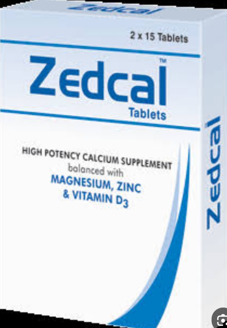
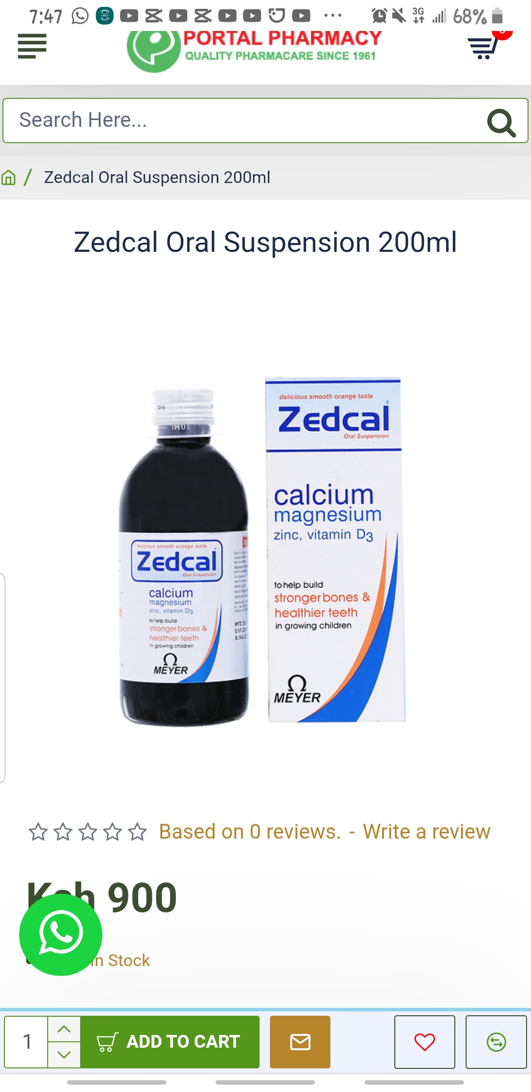
In the clinical management of Hypophosphatemic Rickets, the primary therapeutic objective is to normalize serum phosphate levels and correct skeletal deformities, which necessitates a structured approach to mineral and hormonal supplementation. While the pathophysiology of this condition is distinct from nutritional rickets, the adjunctive use of Vitamin D analogs (e.g., Calcitriol) / نظائر فيتامين د (مثل: كالسيتريول) Standard and broader Vitamin D Analogs / نظائر فيتامين د Standard is often essential to manage secondary hyperparathyroidism and optimize bone mineralization. Clinicians may also integrate Vitamin D supplements / مكملات فيتامين د Standard, Vitamin D3 / فيتامين د3 Varies (e.g., 1000 IU, 5000 IU, 50000 IU), Vitamin D3 (Cholecalciferol) / فيتامين د3 (كولي كالسيفيرول) 50,000 IU, and Vitamin D3 50,000 IU / فيتامين د3 50,000 وحدة دولية 50,000 IU to ensure adequate vitamin D status, alongside calcium-fortified therapies such as Calcimed D3 Effervescent Tablets / أقراص كالسي ميد د3 الفوارة 600 mg Calcium / 400 IU Cholecalciferol, [Calcitron (Calcium Carbonate + Vitamin D3) / كالسيترون (كربونات الكالسيوم + فيتامين د3) Variable (e.g., Calcium Carbonate 500mg, Vitamin D](https
Treatment & Management Options
Recommended Medications