Clinical Assessment & Protocol
Typical Presentation (HPI)
EN: Patient presents with chronic, progressive left shoulder pain localized to the glenohumeral joint. Symptoms are exacerbated by overhead activity, reaching, and internal rotation. Patient reports morning stiffness lasting <30 minutes, mechanical symptoms including crepitus, and progressive loss of range of motion. Conservative measures including NSAIDs and activity modification have provided minimal relief. AR: يراجع المريض بشكوى ألم مزمن ومترقٍ في الكتف الأيسر متمركز في المفصل الحقاني العضدي. تتفاقم الأعراض مع الأنشطة فوق مستوى الرأس، والوصول للأشياء، والدوران الداخلي. يبلغ المريض عن تيبس صباحي يستمر أقل من 30 دقيقة، وأعراض ميكانيكية تشمل فرقعة المفصل، وفقدان تدريجي في مدى الحركة. لم توفر التدابير التحفظية بما في ذلك مضادات الالتهاب غير الستيرويدية وتعديل الأنشطة سوى تحسن طفيف.
General Examination
EN: Inspection of the left shoulder reveals atrophy of the supraspinatus and infraspinatus fossae. Palpation demonstrates tenderness over the glenohumeral joint line and anterior aspect. Range of motion is significantly restricted in both active and passive planes, particularly abduction and external rotation. Crepitus is palpable upon passive circumduction. Neurovascular status is intact distally. AR: يكشف فحص الكتف الأيسر عن ضمور في حفرتي العضلة فوق الشوكة وتحت الشوكة. يظهر الجس وجود إيلام عند خط المفصل الحقاني العضدي والجانب الأمامي. مدى الحركة مقيد بشكل ملحوظ في المستويين النشط والسلبي، وخاصة في الإبعاد والدوران الخارجي. يمكن الشعور بفرقعة عند إجراء الدوران السلبي للمفصل. الحالة العصبية الوعائية سليمة في الأطراف البعيدة.
Treatment Protocol
EN: Plan includes initiation of physical therapy focusing on range of motion and periscapular strengthening. Recommended activity modification to avoid provocative overhead maneuvers. Prescribed NSAIDs for symptom management. Discussed potential for intra-articular corticosteroid or viscosupplementation injection if symptoms persist. AR: تشمل الخطة البدء بالعلاج الطبيعي مع التركيز على مدى الحركة وتقوية العضلات المحيطة بالكتف. يوصى بتعديل الأنشطة لتجنب الحركات المستفزة فوق مستوى الرأس. تم وصف مضادات الالتهاب غير الستيرويدية للتحكم في الأعراض. تمت مناقشة إمكانية حقن الكورتيكوستيرويد داخل المفصل أو حقن المادة الزلالية (Viscosupplementation) في حال استمرار الأعراض.
Patient Education
EN: Glenohumeral osteoarthritis is a degenerative condition involving the wearing down of articular cartilage. Management focuses on symptom control and maintaining joint function. Avoid heavy lifting and repetitive overhead reaching. Adherence to a home exercise program is essential to prevent further stiffness. Seek medical attention if you experience sudden worsening of pain, night pain, or neurological symptoms. AR: التهاب المفاصل الحقاني العضدي هو حالة تنكسية تتضمن تآكل الغضروف المفصلي. يركز العلاج على التحكم في الأعراض والحفاظ على وظيفة المفصل. تجنب رفع الأثقال والوصول المتكرر للأشياء فوق مستوى الرأس. الالتزام ببرنامج التمارين المنزلية ضروري لمنع المزيد من التيبس. يجب مراجعة الطبيب في حال حدوث تفاقم مفاجئ في الألم، أو ألم ليلي، أو أعراض عصبية.
Systemic & Specialized Examinations
EN: Distal neurovascular status intact globally. AR: الحالة العصبية والوعائية الطرفية سليمة تماماً.
Orthopedic & Trauma Assessments
EN: Insidious degenerative wear and tear. No acute trauma. AR: تآكل تنكسي تدريجي. لا توجد صدمة حادة.
EN: Antalgic gait. Reduced stance phase on the affected side. Trendelenburg or varus thrust may be present. AR: مشية متألمة. قصر في مرحلة الوقوف على الجانب المصاب. قد يوجد اندفاع تقوسي أو علامة ترندلينبورغ.
EN: Moderate chronic joint effusion/thickening. Obvious malalignment in the coronal plane. Mild surrounding muscle atrophy. AR: انصباب/تسمك مفصلي مزمن. سوء محاذاة واضح. ضمور خفيف في العضلات المحيطة.
EN: Grind tests (Patellar/FABER) strongly positive. Ligament tests negative. AR: اختبارات الطحن (مثل FABER) إيجابية بقوة. اختبارات الأربطة سلبية.
EN: 4/5 strength in proximal muscles due to pain inhibition. Distal strength 5/5. AR: قوة 4/5 في العضلات القريبة بسبب تثبيط الألم. القوة الطرفية 5/5.
EN: Sensation intact to light touch in all dermatomes. AR: الإحساس سليم للمس الخفيف في جميع التوزيعات العصبية.
EN: 2+ symmetric deep tendon reflexes. AR: المنعكسات العميقة 2+ ومتماثلة.
EN: DP and PT pulses 2+ bounding. Capillary refill < 2 seconds. AR: نبضات القدم 2+ قوية. عودة امتلاء الشعيرات < ثانيتين.
Comprehensive Clinical Guide: Glenohumeral Osteoarthritis (Left Shoulder)
1. Introduction and Clinical Overview
Glenohumeral osteoarthritis (GHOA) represents the end-stage clinical manifestation of progressive articular cartilage degradation within the glenohumeral joint. While the shoulder complex is comprised of four distinct joints, the glenohumeral joint—the primary articulation between the humeral head and the glenoid fossa—is the most susceptible to degenerative changes. When localized to the left shoulder, it presents a unique clinical challenge, often impacting the patient’s dominant or non-dominant activities of daily living (ADLs), necessitating a structured, evidence-based approach to diagnosis and management.
GHOA is characterized by the irreversible thinning of the hyaline cartilage, the formation of marginal osteophytes, subchondral bone sclerosis, and the development of subchondral cysts. Unlike the hip or knee, which are primary weight-bearing joints, the shoulder is a non-weight-bearing joint governed by complex muscular constraints. Consequently, the clinical presentation and progression of GHOA differ significantly from lower extremity arthropathies.
2. Deep-Dive: Etiology and Pathophysiology
Etiology
GHOA can be classified as primary (idiopathic) or secondary. Primary GHOA is less common in the shoulder than in the hip or knee, often associated with age-related senescence. Secondary GHOA, however, is the more frequent clinical encounter:
- Post-Traumatic: Resulting from previous fractures (proximal humerus), recurrent glenohumeral dislocations, or labral tears.
- Rotator Cuff Arthropathy: Massive, chronic rotator cuff tears leading to superior migration of the humeral head, causing eccentric loading and cartilage wear.
- Inflammatory Arthritides: Rheumatoid arthritis, ankylosing spondylitis, or crystal-induced arthropathy (CPPD).
- Avascular Necrosis (AVN): Interruption of the humeral head blood supply due to steroid use, alcohol abuse, or sickle cell disease.
- Infection: Septic arthritis sequelae.
Pathophysiology
The degradation follows a specific biological cascade:
1. Chondrocyte Activation: Stress induces chondrocytes to release pro-inflammatory cytokines (IL-1, TNF-alpha).
2. Matrix Degradation: Matrix metalloproteinases (MMPs) break down the collagen II framework and proteoglycans.
3. Subchondral Remodeling: Loss of cartilage increases stress on the subchondral bone, leading to bone marrow lesions (BMLs) and sclerosis.
4. Osteophyte Formation: Attempted joint stabilization via peripheral bone growth, which further restricts range of motion (ROM).
3. Clinical Staging and Grading
The most widely utilized system for staging GHOA is the Samilson-Prieto Classification, which focuses on the size of the osteophytes on an anteroposterior (AP) radiograph.
| Grade | Description |
|---|---|
| Normal | No osteophytes; joint space > 2mm. |
| Mild | Osteophytes < 3mm. |
| Moderate | Osteophytes 3–7mm; moderate joint space narrowing. |
| Severe | Osteophytes > 7mm; significant joint space obliteration. |
4. Standard Clinical Presentation
Patients with left-sided GHOA typically present with a constellation of symptoms that worsen over time:
- Pain: Deep, aching pain localized to the posterior or lateral aspect of the shoulder. Pain is often worse at night and during activity.
- Stiffness: A progressive loss of glenohumeral motion. Patients often notice difficulty with internal rotation (e.g., reaching into a back pocket) or reaching overhead.
- Crepitus: Palpable or audible grinding during active or passive range of motion.
- Functional Limitations: Inability to perform ADLs, such as dressing, grooming, or reaching for items on high shelves.
- Muscle Atrophy: Chronic disuse may lead to visible wasting of the infraspinatus and supraspinatus muscles.
5. Diagnostic Methodology
Physical Examination
- Inspection: Look for muscle wasting (deltoid, rotator cuff).
- Palpation: Tenderness at the joint line or over the biceps tendon.
- ROM Assessment: Compare to the unaffected side. Passive ROM is typically restricted in both capsular patterns (external rotation, abduction, internal rotation).
- Provocative Testing: The "Grind Test" (axial loading of the humerus while rotating) is often positive for pain and crepitus.
Imaging
- Radiography (Gold Standard):
- Grashey View: True AP view of the glenohumeral joint.
- Axillary View: Essential for evaluating the centering of the humeral head and posterior glenoid wear.
- Scapular Y View: To assess the acromiohumeral interval (important for ruling out rotator cuff arthropathy).
- MRI (Magnetic Resonance Imaging): Indicated if there is suspicion of associated soft tissue pathology, such as a rotator cuff tear or labral pathology.
- CT Scan: Reserved for pre-operative planning to assess glenoid version and bone stock.
6. Differential Diagnosis
It is critical to distinguish GHOA from other shoulder pathologies that may mimic its presentation:
* Adhesive Capsulitis (Frozen Shoulder): Characterized by global loss of ROM; typically self-limiting.
* Rotator Cuff Tendinopathy: Pain is usually arc-specific; ROM is often preserved unless a full-thickness tear is present.
* Cervical Radiculopathy: Referred pain from the C5-C6 nerve roots; neurologic deficits (numbness/tingling) are common.
* Subacromial Impingement Syndrome: Pain is primarily with overhead activity; lack of profound joint space narrowing.
7. Management Strategies
Non-Operative (First-Line)
- Activity Modification: Avoiding high-impact or overhead activities that exacerbate symptoms.
- Physical Therapy: Focus on rotator cuff strengthening, scapular stabilization, and maintaining capsular flexibility.
- Pharmacology: NSAIDs (as tolerated), topical analgesics, or occasional corticosteroid injections for acute flares.
- Viscosupplementation: Intra-articular hyaluronic acid injections (evidence is mixed but often considered).
Operative (Surgical)
- Arthroscopic Debridement: Indicated for mild cases with mechanical symptoms (loose bodies).
- Total Shoulder Arthroplasty (TSA): The gold standard for end-stage GHOA with an intact rotator cuff.
- Reverse Total Shoulder Arthroplasty (rTSA): Indicated if there is significant rotator cuff deficiency (cuff tear arthropathy).
- Hemiarthroplasty: Reserved for younger patients with limited glenoid wear.
8. Risks, Side Effects, and Contraindications
- NSAIDs: Risks include gastrointestinal bleeding, renal impairment, and cardiovascular events.
- Corticosteroid Injections: Potential for infection, cartilage toxicity with repetitive use, and localized skin depigmentation.
- Surgical Risks:
- Infection: Risk of periprosthetic joint infection.
- Instability: Component loosening or dislocation.
- Nerve Injury: Risk to the axillary nerve.
- Contraindications for TSA: Active infection, deltoid paralysis, or severe glenoid bone loss that prevents component fixation.
9. Long-Term Prognosis
The prognosis for GHOA is largely dependent on the stage at diagnosis and the patient’s activity requirements. While non-operative measures can manage symptoms for years, the condition is progressive. Surgical intervention, particularly TSA, has a high success rate, with over 90% of patients reporting significant pain relief and improved function at 10-year follow-ups. However, patients must be counseled that even with successful surgery, the shoulder will not return to "pre-injury" performance levels, and heavy overhead lifting should be avoided to prolong implant longevity.
10. Frequently Asked Questions (FAQ)
1. Is GHOA in the left shoulder more common than the right?
No, it is generally bilateral or unilateral based on injury history. If the left shoulder is non-dominant, patients may ignore symptoms longer, leading to a later-stage presentation.
2. Does diet play a role in GHOA?
While no specific diet cures arthritis, an anti-inflammatory diet (rich in Omega-3s) may help manage systemic inflammation.
3. Can I still exercise with GHOA?
Yes, low-impact exercise is encouraged. Swimming and cycling are excellent, while heavy overhead lifting should be avoided.
4. How often can I get a cortisone injection?
Most clinicians limit injections to 3–4 per year due to concerns regarding cartilage degradation.
5. Is surgery my only option for "severe" grade?
Surgery is the definitive treatment for severe grade, but if the patient can tolerate the pain and has minimal functional demand, conservative management can be continued indefinitely.
6. What is the difference between TSA and rTSA?
TSA replaces the ball and socket in the anatomical position. rTSA reverses the anatomy, placing the ball on the glenoid side, which compensates for a non-functional rotator cuff.
7. How long is the recovery from shoulder replacement?
Full recovery typically takes 6–12 months, involving an initial period of immobilization followed by intensive physical therapy.
8. Can cold weather affect my shoulder pain?
Yes. Changes in barometric pressure can cause expansion of joint tissues, leading to increased pain in arthritic joints.
9. Is GHOA hereditary?
Genetics play a role in primary osteoarthritis, particularly in the development of cartilage susceptibility.
10. Can physical therapy make the pain worse?
Initially, some discomfort is normal as range of motion is challenged, but sharp, lingering pain is a sign that the intensity or type of exercise should be adjusted.
Summary Table: Clinical Decision Making
| Feature | Mild GHOA | Severe GHOA |
|---|---|---|
| Primary Treatment | PT, NSAIDs, Modification | Surgical Consultation |
| Imaging Finding | Small osteophytes | Bone-on-bone / Large osteophytes |
| Goal | Symptom Management | Pain relief & Function restoration |
| Follow-up | 6–12 months | Pre-surgical planning / Post-op |
Disclaimer: This guide is intended for educational purposes for clinical professionals and medical students. It does not replace professional medical judgment or direct patient care protocols. Always consult with an orthopedic specialist for individual case management.
Related Clinical Integration
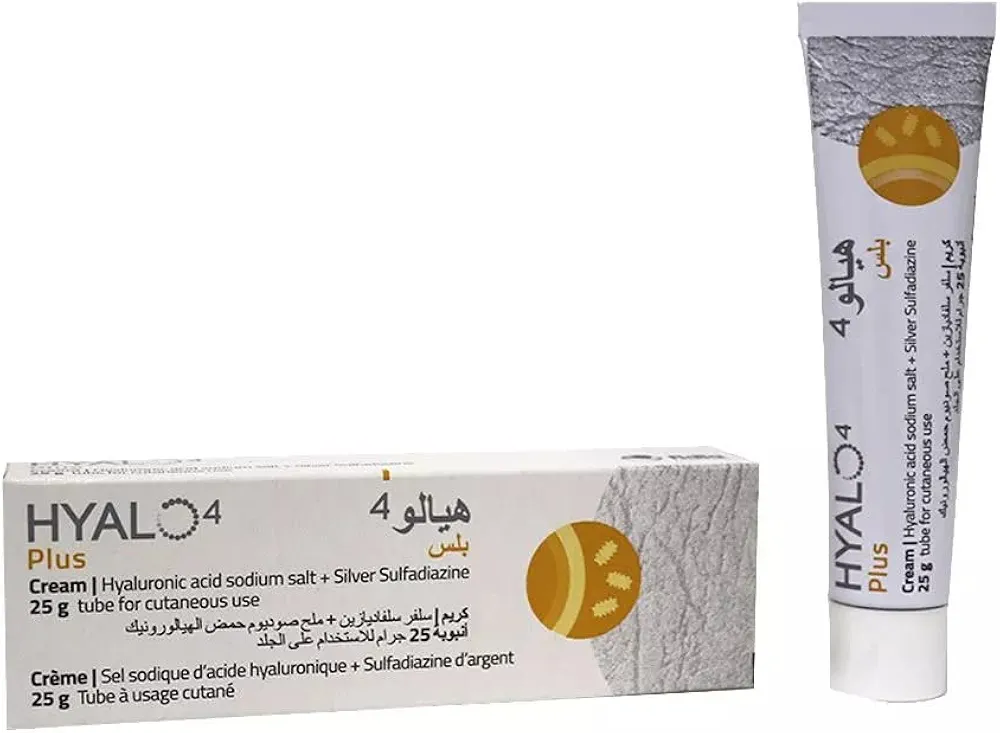
In the management of Glenohumeral Osteoarthritis, Left Shoulder, a comprehensive clinical approach integrates pharmacological, surgical, and educational resources to optimize patient outcomes. Initial conservative therapy often involves anti-inflammatory support with Celcox / سيلكوكس 100mg and topical care using Hyalo4 plus cream / هيالو 4 بلس كريم 0.2% / 1%, while physical comfort is maintained through the use of a Simple Shoulder Sling (UltraSling) / حمالة كتف بسيطة (ألتراسلينغ) (الأطراف الصناعية والجبائر التقويمية). When surgical intervention becomes necessary, specialized equipment such as Trephine Reamer Sets / مجموعات موسعات الثقب (تريفين) and Oscillating Bone Saw Blade (Wide, Narrow, Deep Cut) / شفرة منشار عظمي متذبذب (عريض، ضيق، قطع عميق) are essential for procedures like Reverse Total Shoulder Arthroplasty (rTSA) / رأب مفصل الكتف الكلي المعكوس (rTSA) (عملية كبرى في غرف العمليات) or Cemented Hemiarthroplasty for Pathologic Fracture / رأب نصف المفصل الملحوم لكسر مرضي (عملية كبرى في غرف العمليات). While specialized hardware like [Distal Femoral Replacement (Tumor Prosthesis) / استبدال الفخذ البعيد (طرف صناعي للأورام)](https://yemenhealthos.com/ar/clinic/instruments/distal-femoral-replacement-tumor-prosthesis-f59a7