Clinical Assessment & Protocol
Typical Presentation (HPI)
EN: Patient presents with persistent burning pain, hyperalgesia, and vasomotor changes following a deep laceration of the median nerve. AR: يعاني المريض من ألم حارق مستمر، وفرط التألم، وتغيرات وعائية بعد تمزق عميق في العصب المتوسط.
General Examination
EN: Allodynia, trophic changes in skin, localized edema, and sweating abnormalities in the affected hand. AR: فرط الحس اللمسي، تغيرات تغذوية في الجلد، وذمة موضعية، واضطرابات في التعرق في اليد المصابة.
Treatment Protocol
EN: Multidisciplinary approach including physical therapy, sympathetic nerve blocks, and gabapentinoids. AR: نهج متعدد التخصصات يشمل العلاج الطبيعي، وحقن الأعصاب الودية، وأدوية الغابابنتينويد.
Patient Education
EN: Early mobilization and desensitization exercises are crucial for long-term functional recovery. AR: تعتبر الحركة المبكرة وتمارين إزالة التحسس ضرورية للتعافي الوظيفي على المدى الطويل.
Systemic & Specialized Examinations
EN: S1, S2 present. No murmurs. AR: صوتا القلب الأول والثاني طبيعيان. لا توجد نفخات.
EN: Lungs clear to auscultation. AR: الرئتان صافيتان عند التسمع.
EN: Abdomen soft, non-tender. AR: البطن لين ولا يوجد ألم.
EN: Alert, oriented x3. No focal deficits. AR: المريض واعي ومدرك. لا يوجد عجز عصبي بؤري.
EN: Unremarkable or not routinely indicated. AR: طبيعي أو غير مطلوب روتينياً.
EN: Unremarkable or not routinely indicated. AR: طبيعي أو غير مطلوب روتينياً.
EN: Unremarkable or not routinely indicated. AR: طبيعي أو غير مطلوب روتينياً.
EN: Unremarkable or not routinely indicated. AR: طبيعي أو غير مطلوب روتينياً.
EN: Unremarkable or not routinely indicated. AR: طبيعي أو غير مطلوب روتينياً.
Orthopedic & Trauma Assessments
EN: Unremarkable or not routinely indicated. AR: طبيعي أو غير مطلوب روتينياً.
EN: Unremarkable or not routinely indicated. AR: طبيعي أو غير مطلوب روتينياً.
Comprehensive Clinical Guide: Complex Regional Pain Syndrome Type II (Causalgia)
1. Introduction and Clinical Overview
Complex Regional Pain Syndrome Type II (CRPS-II), historically referred to as "Causalgia," represents a severe, chronic neuropathic pain condition that follows a distinct nerve injury. Unlike CRPS-I (Reflex Sympathetic Dystrophy), where no specific nerve lesion can be identified, CRPS-II is characterized by the presence of a definable, traumatic injury to a peripheral nerve.
This condition is a debilitating neuro-inflammatory disorder. It is characterized by persistent, intense pain that is disproportionate to the inciting event, often accompanied by sensory, autonomic, sudomotor, and motor disturbances. Given the complex nature of its pathophysiology—which involves the central nervous system (CNS), the peripheral nervous system (PNS), and the immune system—CRPS-II remains one of the most challenging diagnoses in orthopedic and pain medicine.
2. Etiology and Pathophysiology
The pathophysiology of CRPS-II is multifactorial, involving a "perfect storm" of maladaptive neuroplasticity and persistent inflammation.
The Mechanism of Injury
CRPS-II is triggered by damage to a peripheral nerve (e.g., crush injury, laceration, surgery, or prolonged compression). This trauma initiates a cascade of events that sensitizes both peripheral and central pain pathways.
Key Pathophysiological Pillars:
- Peripheral Sensitization: Damaged nerve fibers (nociceptors) increase their firing rate, often manifesting as spontaneous ectopic discharges. This lowers the threshold for pain, turning innocuous stimuli into painful ones (allodynia).
- Central Sensitization: Sustained peripheral input leads to hyperexcitability of the dorsal horn neurons in the spinal cord. This "wind-up" effect creates a state where the CNS amplifies pain signals even in the absence of ongoing peripheral tissue damage.
- Neurogenic Inflammation: Activation of sensory neurons releases neuropeptides such as Substance P and Calcitonin Gene-Related Peptide (CGRP). This causes localized vasodilation, plasma extravasation, and the recruitment of inflammatory cytokines (IL-1, IL-6, TNF-alpha).
- Sympathetic-Afferent Coupling: In CRPS-II, adrenergic receptors (alpha-adrenoceptors) may develop on the surface of nociceptive fibers. Consequently, norepinephrine released by the sympathetic nervous system can directly stimulate pain fibers, linking the sympathetic nervous system to the pain experience.
3. Clinical Staging and Presentation
CRPS-II does not follow a strict linear progression, but clinicians generally categorize the disorder into three clinical stages based on temporal and symptomatic evolution.
| Stage | Timing (Approx.) | Clinical Characteristics |
|---|---|---|
| Stage I (Acute) | 0–3 Months | Severe burning pain, localized edema, hyperhidrosis, increased hair/nail growth, vasodilation. |
| Stage II (Dystrophic) | 3–6 Months | Pain intensifies, edema becomes indurated (hardened), skin becomes thin/cyanotic, muscle wasting begins. |
| Stage III (Atrophic) | 6+ Months | Irreversible changes, skin becomes cold/pale, severe muscle atrophy, joint contractures, bone demineralization. |
Standard Symptomatic Presentation
- Sensory: Hyperalgesia (increased sensitivity to pain) and Allodynia (pain from non-painful stimuli like light touch or wind).
- Vasomotor/Sudomotor: Temperature asymmetry between limbs, color changes (mottling, cyanosis), and excessive or absent sweating.
- Motor/Trophic: Reduced range of motion, tremors, dystonia, or weakness. Trophic changes include brittle nails, sparse hair growth, and thinning skin.
4. Differential Diagnosis
Distinguishing CRPS-II from other neuropathic conditions is critical, as treatment protocols vary significantly.
- Peripheral Neuropathy: Usually symmetrical and distal (e.g., diabetic neuropathy), whereas CRPS-II is regional and often unilateral.
- Vascular Insufficiency: Peripheral Arterial Disease (PAD) may cause pain, but lacks the neurological sensory disturbances (allodynia) typical of CRPS.
- Infection/Cellulitis: Can mimic the redness and heat of early CRPS, but lacks the chronic, disproportionate pain profile.
- Thoracic Outlet Syndrome (TOS): May present with limb pain and vascular changes, but is localized to the brachial plexus distribution.
- Factitious Disorder: Must be considered if the clinical findings are inconsistent with anatomical distribution or if there is secondary gain involved.
5. Diagnostic Criteria (Budapest Criteria)
The Budapest Criteria are the gold standard for clinical diagnosis. To meet the criteria, the patient must have:
- Continuing pain which is disproportionate to any inciting event.
- At least one symptom in three of the four following categories:
- Sensory: Hyperesthesia or Allodynia.
- Vasomotor: Temperature asymmetry or color changes.
- Sudomotor/Edema: Edema or sweating changes.
- Motor/Trophic: Decreased ROM, motor dysfunction (tremor, dystonia), or trophic changes (hair/nail/skin).
- At least one sign (observed on exam) in two or more of the same categories.
- No other diagnosis that better explains the signs and symptoms.
6. Diagnostic Testing and Imaging
There is no single "gold standard" test for CRPS-II. Diagnosis remains primarily clinical, supported by exclusionary testing.
- Electromyography (EMG) / Nerve Conduction Studies (NCS): Essential to confirm the underlying nerve injury required for a Type II diagnosis.
- Tri-phase Bone Scan: Can detect increased uptake in the affected region, indicating regional bone metabolic changes (common in early stages).
- MRI: Useful for identifying structural nerve damage, muscle atrophy, or bone marrow edema.
- Sympathetic Blockade: Diagnostic (and sometimes therapeutic) injection of local anesthetic into the stellate ganglion (for upper limbs) or lumbar sympathetic chain (for lower limbs). If pain resolves, it suggests a sympathetic component.
7. Risks, Contraindications, and Management Considerations
Risks of Untreated CRPS-II
- Permanent physical disability.
- Severe psychological distress (depression, anxiety, suicidal ideation).
- Irreversible joint contractures and muscle atrophy.
Contraindications in Management
- Avoid unnecessary surgery: Surgical intervention in an area affected by active CRPS can lead to a "flare" or exacerbation of symptoms.
- Avoid aggressive physical therapy: Pushing through severe pain can trigger central sensitization; therapy must be graded and desensitization-focused.
Multi-Modal Treatment Approach
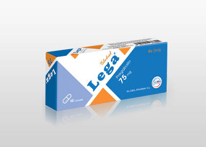
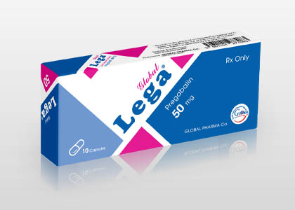
- Pharmacotherapy: Gabapentinoids (Gabapentin, Pregabalin), Tricyclic Antidepressants (Amitriptyline), and NMDA antagonists (Ketamine infusions).
- Interventional: Sympathetic nerve blocks, spinal cord stimulation (SCS), and intrathecal drug delivery systems.
- Psychological: Cognitive Behavioral Therapy (CBT) to manage the chronic pain cycle.
- Physical/Occupational Therapy: Graded motor imagery (GMI), mirror therapy, and desensitization techniques.
8. Long-Term Prognosis
The prognosis for CRPS-II is highly variable. Early diagnosis (within the first 3-6 months) is the strongest predictor of a positive outcome. If the condition progresses to Stage III, permanent structural changes often occur, making the focus shift from "cure" to "functional restoration and pain management."
Many patients achieve significant symptom reduction with multidisciplinary care, though a subset of patients may experience lifelong pain. Success is measured by the ability to engage in activities of daily living (ADLs) and improved quality of life rather than total pain eradication.
9. Frequently Asked Questions (FAQ)
1. Is CRPS-II the same as Reflex Sympathetic Dystrophy (RSD)?
No. CRPS-I is RSD (no confirmed nerve injury). CRPS-II is Causalgia (confirmed nerve injury).
2. Can CRPS-II spread to other limbs?
Yes, "spread" can occur, usually to the contralateral limb or an adjacent region, likely due to central nervous system sensitization.
3. Does a normal MRI rule out CRPS-II?
No. CRPS-II is a clinical diagnosis. Imaging is used to rule out other pathology, not to confirm CRPS.
4. Are there genetic predispositions to CRPS-II?
Some research suggests a genetic predisposition involving inflammatory and HLA-related markers, though it is not strictly hereditary.
5. Why is the pain so much worse than the injury?
The pain is amplified by the nervous system. The injury acts as a "trigger," but the CNS keeps the "alarm" ringing long after the tissue heals.
6. What is the role of Mirror Therapy?
Mirror therapy tricks the brain into thinking the affected limb is moving painlessly, which can help reorganize the cortical maps of the brain and reduce pain.
7. Is surgery always contraindicated?
Not always, but it is high-risk. If surgery is necessary, it is often performed under a "CRPS protocol" involving sympathetic blocks to minimize flare-ups.
8. How effective is Spinal Cord Stimulation (SCS)?
SCS is highly effective for patients who fail conservative management. It masks pain signals by sending electrical impulses to the spinal cord.
9. Can stress trigger a CRPS-II flare?
Yes. The sympathetic nervous system is linked to the stress response, which can exacerbate the pain and vasomotor symptoms of CRPS.
10. Does CRPS-II ever go away?
Some patients achieve remission, especially with early intervention. For others, it becomes a chronic condition that requires lifelong management.
10. Conclusion for Clinical Practitioners
Managing CRPS-II requires a compassionate, multidisciplinary approach. The medical professional must validate the patient’s experience, as the "invisible" nature of the pain often leads to significant psychological distress. By focusing on early diagnosis, aggressive desensitization, and a collaborative team approach involving pain specialists, physical therapists, and psychologists, the practitioner can significantly improve the patient's functional trajectory and overall quality of life.
Disclaimer: This guide is intended for medical informational purposes only and does not constitute formal medical advice. Clinical decisions should be based on institutional protocols and individual patient assessment.
Treatment & Management Options
Recommended Medications