Clinical Assessment & Protocol
Typical Presentation (HPI)
EN: Patient presents with chronic, progressive pain localized to the left thumb base. Symptoms are exacerbated by pinch and grasp activities, including opening jars and turning keys. Patient reports morning stiffness and occasional swelling at the CMC joint. No history of acute trauma. Conservative measures including NSAIDs and splinting have provided [partial/minimal/no] relief. AR: يعاني المريض من ألم مزمن ومتزايد في قاعدة الإبهام الأيسر. تزداد حدة الأعراض مع أنشطة القرص والقبض، مثل فتح العلب أو تدوير المفاتيح. يشكو المريض من تيبس صباحي وتورم متقطع في المفصل الرسغي السنعي. لا يوجد تاريخ لإصابة حادة. الإجراءات التحفظية بما في ذلك مضادات الالتهاب غير الستيرويدية والجبيرة قدمت تحسناً [جزئياً/طفيفاً/معدوماً].
General Examination
EN: Examination of the left hand reveals tenderness to palpation at the trapeziometacarpal (TMC) joint. Grind test is positive, eliciting pain and crepitus. Mild prominence/subluxation noted at the base of the first metacarpal. Range of motion shows restricted thumb abduction and adduction secondary to pain. Neurovascular status is intact distally. AR: كشف فحص اليد اليسرى عن وجود ألم عند الجس في المفصل الرسغي السنعي. اختبار الطحن (Grind test) إيجابي، مما يسبب ألماً وفرقعة. لوحظ بروز/خلع جزئي طفيف في قاعدة المشط الأول. يظهر نطاق الحركة تقيداً في تبعيد وتقريب الإبهام بسبب الألم. الحالة العصبية الوعائية سليمة في الأطراف.
Treatment Protocol
EN: Plan includes: 1. Activity modification and avoidance of repetitive pinch maneuvers. 2. Thumb spica splinting for stabilization. 3. Trial of topical/oral NSAIDs. 4. Consider corticosteroid injection for symptomatic relief. 5. Discuss surgical options (e.g., trapeziectomy with ligament reconstruction) if conservative management fails. AR: تشمل الخطة: 1. تعديل الأنشطة وتجنب حركات القرص المتكررة. 2. استخدام جبيرة الإبهام للتثبيت. 3. تجربة مضادات الالتهاب غير الستيرويدية الموضعية أو الفموية. 4. النظر في حقن الكورتيكوستيرويد لتخفيف الأعراض. 5. مناقشة الخيارات الجراحية (مثل استئصال العظم المربعي مع إعادة بناء الأربطة) في حال فشل العلاج التحفظي.
Patient Education
EN: Basal joint arthritis is wear-and-tear of the cartilage at the base of your thumb. To manage symptoms, wear your splint during heavy activities, apply ice for 15 minutes after use, and perform gentle range-of-motion exercises as directed. Avoid activities that cause sharp pain. Contact the clinic if you experience increased numbness, tingling, or worsening weakness. AR: التهاب مفصل قاعدة الإبهام هو تآكل في الغضروف الموجود في قاعدة إبهامك. للتحكم في الأعراض، ارتدِ الجبيرة أثناء الأنشطة الشاقة، واستخدم الثلج لمدة 15 دقيقة بعد الاستخدام، وقم بتمارين نطاق الحركة اللطيفة حسب التوجيهات. تجنب الأنشطة التي تسبب ألماً حاداً. اتصل بالعيادة إذا شعرت بزيادة في التنميل أو الوخز أو ضعف متزايد.
Systemic & Specialized Examinations
EN: Intact distally. AR: سليم طرفياً.
Orthopedic & Trauma Assessments
EN: Idiopathic inflammatory cascade or hematogenous bacterial seeding. AR: سلسلة التهابات مناعية أو انتشار بكتيري عبر الدم.
EN: Non-ambulatory due to severe joint pain on loading. AR: غير قادر على المشي بسبب الألم الشديد عند تحميل الوزن.
EN: Joint is markedly erythematous, warm, tense, and visibly swollen. Loss of skin wrinkles over the joint. AR: المفصل شديد الاحمرار، دافئ، مشدود، ومتورم بوضوح. فقدان تجاعيد الجلد فوق المفصل.
EN: N/A. Clinical picture dominates. AR: لا ينطبق. الصورة السريرية هي السائدة.
EN: Pseudoparalysis. AR: شلل كاذب.
EN: Intact. AR: سليم.
EN: Deferred. AR: مؤجل.
EN: 2+ symmetric. AR: 2+ متماثلة.
Comprehensive Clinical Guide: Basal Joint Arthritis of the Left Hand
1. Introduction and Clinical Overview
Basal joint arthritis, medically referred to as Trapeziometacarpal (TMC) joint arthritis, represents one of the most prevalent and debilitating degenerative conditions of the upper extremity. It specifically involves the articulation between the trapezium (a carpal bone in the wrist) and the base of the first metacarpal (the thumb).
The left hand, often the non-dominant hand in the general population, is frequently subjected to compensatory stress, particularly in individuals with pre-existing injuries or those who undergo repetitive tasks. As the thumb is responsible for approximately 40% to 50% of total hand function—primarily due to its unique ability to perform opposition—degeneration of the basal joint significantly compromises pinch strength, grip stability, and activities of daily living (ADLs).
This guide provides an exhaustive clinical breakdown of the condition, intended for medical professionals and clinical specialists managing patients with this diagnosis.
2. Etiology and Pathophysiology
The Biomechanics of the TMC Joint
The TMC joint is a saddle-shaped (sellar) joint. This configuration allows for complex multi-axial movement, including flexion, extension, abduction, adduction, and circumduction. However, this high degree of mobility comes at the cost of stability.
Primary Etiological Factors
- Age-Related Degeneration: The most common cause. Cumulative wear and tear on the articular cartilage leads to thinning and eventual bone-on-bone contact.
- Ligamentous Laxity: Chronic attenuation of the anterior oblique ligament (AOL), also known as the "beak ligament," which is the primary stabilizer of the TMC joint.
- Hormonal Influences: The condition is significantly more prevalent in post-menopausal women, suggesting a potential role for estrogen receptors in the maintenance of joint cartilage and ligament integrity.
- Anatomical Variations: Abnormal joint geometry or prior trauma (e.g., intra-articular fractures) can accelerate degenerative changes.
The Pathological Cascade
As the articular cartilage degrades, the joint space narrows. The loss of the AOL causes subluxation of the metacarpal base in a dorsal and radial direction. This subluxation creates a vicious cycle: the joint becomes incongruent, leading to uneven pressure distribution, further cartilage erosion, and the formation of osteophytes (bone spurs) at the joint margins.
3. Clinical Staging and Grading: The Eaton-Littler Classification
To standardize the assessment of left-hand basal joint arthritis, clinicians utilize the Eaton-Littler classification system, which categorizes the disease based on radiographic findings.
| Stage | Radiographic Description |
|---|---|
| Stage I | Normal joint space or slight widening due to effusion; no osteophytes. |
| Stage II | Slight narrowing of the joint space; osteophytes < 2mm. |
| Stage III | Marked narrowing of the joint space; subchondral sclerosis; osteophytes > 2mm. |
| Stage IV | Pan-trapezial arthritis; involvement of the scaphotrapezial joint. |
4. Clinical Presentation and Diagnostic Assessment
Standard Presentation
Patients typically report:
* Localized Pain: Deep, aching pain at the base of the thumb (proximal to the anatomical snuffbox).
* Weakness: Difficulty with "pinch" tasks, such as turning a key, opening jars, or buttoning shirts.
* Morning Stiffness: Symptoms often exacerbate after periods of inactivity.
* Visible Deformity: In advanced stages, a "squared-off" appearance of the thumb base due to metacarpal subluxation and adduction contracture.
Diagnostic Physical Tests
- The Grind Test: The clinician applies axial pressure to the thumb metacarpal while rotating it. A positive result is the reproduction of pain and/or crepitus.
- The Lever Test (Traction-Shift Test): The clinician applies longitudinal traction to the thumb and then shifts the metacarpal base. Pain and hypermobility indicate ligamentous instability.
- Adduction Contracture Assessment: Observing the inability of the patient to fully abduct the thumb away from the index finger (the "Web Space" test).
Imaging Modalities
- Plain Radiographs: Standard views include Posteroanterior (PA), Lateral, and the "Robert’s View" (a true AP view of the TMC joint).
- MRI/Ultrasound: Generally reserved for cases where physical exam is ambiguous or to rule out ligamentous tears (e.g., Gamekeeper’s thumb) if the history suggests acute injury.
5. Differential Diagnosis
It is critical to distinguish basal joint arthritis from other pathology:
* De Quervain’s Tenosynovitis: Inflammation of the tendons in the first dorsal compartment. (Positive Finkelstein’s test).
* Scaphoid Fracture: Often presents with pain in the snuffbox; requires ruling out via imaging if history involves trauma.
* Carpal Tunnel Syndrome: Can cause referred pain or sensory disturbances in the thumb.
* Trigger Thumb: Stenosing tenosynovitis of the flexor pollicis longus.
6. Treatment Modalities and Management
Conservative Management (First-Line)
- Splinting: Use of a thumb spica orthosis to immobilize the TMC joint during periods of high stress.
- Pharmacology: NSAIDs (oral or topical diclofenac) to manage inflammation.
- Injections: Corticosteroid injections into the TMC joint for short-term pain relief. Hyaluronic acid injections are an emerging alternative.
- Hand Therapy: Focus on strengthening the intrinsic muscles of the hand to provide dynamic stability to the joint.
Surgical Intervention
Reserved for patients failing conservative management:
1. Trapeziectomy with Ligament Reconstruction and Tendon Interposition (LRTI): The gold standard for many surgeons.
2. Arthrodesis (Fusion): Fusing the joint to eliminate motion; effective for high-demand, younger patients.
3. Joint Replacement (Arthroplasty): Using prosthetic implants to replace the articular surfaces.
7. Risks and Contraindications
- Infection: Risk associated with any intra-articular injection or surgical procedure.
- Nerve Injury: The superficial radial nerve runs in close proximity to the TMC joint; surgical dissection risks sensory loss.
- CRPS (Complex Regional Pain Syndrome): A rare but severe complication following hand surgery.
- Contraindications for Surgery: Active infection, severe peripheral vascular disease, or unrealistic patient expectations regarding post-operative range of motion.
8. FAQ: Frequently Asked Questions
1. Is basal joint arthritis of the left hand different from the right?
Biologically, no. However, the functional impact differs based on handedness. Left-hand arthritis is often ignored longer by right-handed patients, leading to later-stage presentation.
2. Can diet change the progression of this arthritis?
While no specific diet cures arthritis, an anti-inflammatory diet (rich in Omega-3s) may help manage systemic inflammation.
3. How successful is surgery for the left thumb?
Success rates are high, with the majority of patients reporting significant pain reduction and improved pinch strength within 6 months post-op.
4. Will I lose range of motion after surgery?
Some reduction in thumb abduction is common, but most patients gain far more functional motion by eliminating the pain that previously restricted movement.
5. Are there specific exercises I should avoid?
Avoid heavy, repetitive gripping or twisting motions (like wringing out wet towels) which place maximal torque on the TMC joint.
6. Can I use a brace forever?
Bracing is a tool for flare-ups. Long-term, 24/7 use is discouraged as it may lead to muscle atrophy in the thumb.
7. Is "cracking" the thumb joint bad?
Crepitus (grinding) is a symptom of the arthritis itself, not the cause. However, repetitive intentional cracking provides no clinical benefit.
8. Does cold weather affect the left hand more?
Yes, barometric pressure changes and cold temperatures often cause joint stiffness and increased pain perception in arthritic joints.
9. How long does the recovery take after a trapeziectomy?
Full recovery typically takes 6 to 12 months, with the first 6 weeks involving strict immobilization.
10. Can basal joint arthritis lead to other hand issues?
Yes, untreated TMC arthritis can lead to "swan-neck" deformity of the thumb as the body attempts to compensate for the unstable basal joint.
9. Long-Term Prognosis
The long-term prognosis for patients with basal joint arthritis of the left hand is generally favorable. While the condition is progressive, most patients can be effectively managed with a combination of activity modification, orthotic support, and, if necessary, surgical reconstruction. Early clinical intervention is the key to preventing the adduction contractures that severely limit the hand's functional utility.
Patients should be monitored annually for the development of secondary deformities, and surgeons should emphasize the importance of post-operative hand therapy to ensure optimal functional outcomes. With modern surgical techniques, the prognosis for restoring a pain-free, functional pinch is excellent for the vast majority of patients.
Related Clinical Integration
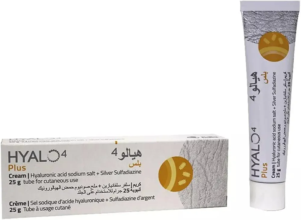
In the management of Basal Joint Arthritis (Thumb), Left Hand, a multidisciplinary clinical approach is essential to optimize patient outcomes, ranging from conservative symptom management to advanced surgical intervention. Initial non-surgical protocols typically involve the use of a Thumb Spica Splint / جبيرة إبهام سبايكا (الأطراف الصناعية والجبائر التقويمية) to provide joint stability, often supplemented by pharmacological therapies such as Celcox / سيلكوكس 100mg, Mediflam D.T / ميديفلام دي تي 50 mg, Meloxicam / ميلوكسيكام 25mg, or topical applications like Hyalo4 plus cream / هيالو 4 بلس كريم 0.2% / 1%. For patients requiring operative management, specialized equipment such as the Bone Rongeur (Leksell) / ملقط عظم (ليكسيل) is utilized in procedures ranging from Thumb Carpometacarpal Joint Arthrodesis: An Intraoperative Masterclass for Basal Joint Arthritis to Thumb Carpometacarpal Joint Arthroplasty: An Intraoperative Masterclass in Resurfacing and Total Joint Replacement. While procedures like [Lateral Ankle Ligament Reconstruction (Brostrom/Modified Brostrom) / إعادة بناء أربطة الكاحل الجانبية (بطريقة بروسترم/بروسترم المعدلة) (عملية كبرى في غرف العمليات)](https://yemenhealthos.com/ar/clinic/medical-procedures/lateral-ankle-ligament-