Clinical Assessment & Protocol
Typical Presentation (HPI)
EN: Patient presents with a [duration] history of [symptom, e.g., joint pain/fatigue/rash], characterized by [quality, e.g., morning stiffness lasting >1 hour]. Symptoms are [improving/worsening] and are associated with [associated symptoms]. AR: يراجع المريض بتاريخ مرضي منذ [المدة] لـ [العرض، مثل: ألم المفاصل/الإرهاق/الطفح الجلدي]، يوصف بأنه [طبيعة العرض، مثل: تيبس صباحي يستمر لأكثر من ساعة]. الأعراض [تتحسن/تسوء] وتترافق مع [الأعراض المصاحبة].
General Examination
EN: Patient is [alert/oriented/distressed]. Vital signs: Temp [temp], BP [bp], HR [hr]. General appearance: [well-appearing/ill-appearing]. No acute distress noted. AR: المريض [واعٍ/مدرك/يعاني من ضيق]. العلامات الحيوية: الحرارة [درجة الحرارة]، ضغط الدم [ضغط الدم]، نبض القلب [النبض]. المظهر العام: [بحالة جيدة/يبدو مريضاً]. لا توجد علامات ضيق حاد.
Treatment Protocol
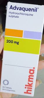
EN: Plan: Initiate [medication name] at [dosage] once daily. Advise patient on [side effects/monitoring]. Follow-up in [duration] for [labs/clinical assessment]. AR: الخطة: البدء بـ [اسم الدواء] بجرعة [الجرعة] مرة يومياً. توجيه المريض بشأن [الآثار الجانبية/المتابعة]. المراجعة بعد [المدة] لـ [الفحوصات المخبرية/التقييم السريري].
Patient Education
EN: Patient educated on the chronic nature of [diagnosis]. Discussed importance of medication adherence, lifestyle modifications, and early reporting of [warning signs]. Provided with educational pamphlets. AR: تم تثقيف المريض حول الطبيعة المزمنة لـ [التشخيص]. تمت مناقشة أهمية الالتزام بالدواء، وتعديلات نمط الحياة، والإبلاغ المبكر عن [علامات التحذير]. تم تزويد المريض بمنشورات تثقيفية.
Orthopedic & Trauma Assessments
EN: Range of motion (ROM) is [full/restricted] in [joints]. Limitations noted in [flexion/extension/rotation]. AR: مدى الحركة (ROM) [كامل/محدود] في [المفاصل]. لوحظت قيود في [الثني/البسط/الدوران].
The Comprehensive Medical Guide to Autoimmune Diseases: Rheumatoid Arthritis and Systemic Lupus Erythematosus
1. Comprehensive Introduction & Overview
Autoimmune diseases represent a complex and often debilitating category of chronic conditions where the body's immune system, which is designed to protect against foreign invaders like bacteria and viruses, mistakenly attacks its own healthy tissues. This fundamental breakdown in self-tolerance leads to inflammation, tissue damage, and a wide array of clinical manifestations affecting virtually any organ system. The impact on patients can range from chronic pain and fatigue to severe organ dysfunction and reduced quality of life.
This guide delves into two prominent examples of systemic autoimmune diseases: Rheumatoid Arthritis (RA) and Systemic Lupus Erythematosus (SLE). While both share the common thread of immune dysregulation, they present with distinct clinical patterns, specific diagnostic markers, and unique challenges in management. Understanding these conditions is crucial for healthcare professionals and patients alike, enabling timely diagnosis, effective treatment strategies, and improved long-term outcomes. Globally, autoimmune diseases affect millions, with a higher prevalence often observed in women, highlighting their significant public health burden.
2. Deep-dive into Technical Specifications / Mechanisms
2.1 Clinical Definition
Autoimmune diseases are characterized by an adaptive immune response directed against self-antigens. This pathology arises from a failure of immune tolerance, a state where the immune system distinguishes between self and non-self. When tolerance is lost, autoreactive T cells and B cells become activated, leading to the production of autoantibodies and pro-inflammatory cytokines that mediate tissue destruction.
- Rheumatoid Arthritis (RA): A chronic, systemic inflammatory disorder primarily affecting the synovial lining of joints, leading to progressive joint destruction, deformity, and functional disability. It is characterized by symmetrical polyarthritis, particularly of the small joints.
- Systemic Lupus Erythematosus (SLE): A chronic, multi-system inflammatory autoimmune disease characterized by the production of numerous autoantibodies, immune complex formation, and widespread inflammation affecting joints, skin, kidneys, brain, heart, lungs, and blood cells. It is often referred to as "the great imitator" due to its variable and diverse clinical presentation.
2.2 Etiology: The Multifactorial Origins
The exact cause of autoimmune diseases is often unknown, but it is understood to be a complex interplay of genetic predisposition and environmental triggers.
- Genetic Predisposition:
- HLA Genes: The human leukocyte antigen (HLA) complex, particularly HLA-DRB1 alleles (e.g., HLA-DRB1*0401, *0404, *0101 for RA; HLA-DRB1*0301 for SLE), are strongly associated with increased risk. These genes play a critical role in presenting antigens to T cells.
- Non-HLA Genes: Numerous other genes contribute to susceptibility, including those involved in immune regulation (e.g., PTPN22, STAT4, TRAF1/C5 in RA; STAT4, IRF5, BLK, PTPN22 in SLE), apoptosis, and clearance of immune complexes.
- Environmental Triggers:
- Infections: Viral (e.g., Epstein-Barr Virus in SLE, parvovirus B19) and bacterial infections are hypothesized to trigger autoimmunity via molecular mimicry (where microbial antigens resemble self-antigens) or bystander activation.
- Smoking: A well-established risk factor for RA, particularly in individuals with genetic predisposition (e.g., HLA-DRB1*0401), potentially by inducing citrullination of proteins in the lungs.
- UV Light Exposure: A significant trigger for SLE flares, particularly skin manifestations and systemic disease activity.
- Dietary Factors/Microbiome: Emerging research suggests the gut microbiome (dysbiosis) may influence immune responses and contribute to autoimmune disease development.
- Toxins/Chemicals: Exposure to certain chemicals (e.g., silica, pesticides) has been linked to increased risk.
- Hormonal Factors:
- Sex Hormones: The higher prevalence of autoimmune diseases in women, particularly during reproductive years, suggests a role for estrogens, which can modulate immune responses. For instance, estrogen can enhance B cell activity.
- Epigenetic Modifications: Changes in gene expression without altering the DNA sequence (e.g., DNA methylation, histone modification) can influence immune cell function and contribute to autoimmunity.
2.3 Pathophysiology: Mechanisms of Self-Attack
The pathogenesis involves a breakdown of immune tolerance at multiple levels, leading to chronic inflammation and tissue damage.
- Loss of Central Tolerance: Defects in the deletion of autoreactive T and B cells in the thymus and bone marrow, respectively.
- Loss of Peripheral Tolerance: Failure of regulatory T cells (Tregs) to suppress autoreactive immune responses in the periphery, or defects in anergy (inactivation) or apoptosis of self-reactive lymphocytes.
- Antigen Presentation: In RA, post-translational modification of proteins (e.g., citrullination) creates novel self-antigens that are presented by HLA-DRB1 molecules to autoreactive T cells. In SLE, nuclear antigens (DNA, histones, Sm, RNP) are released, possibly due to impaired clearance of apoptotic cells, and presented to immune cells.
- B Cell Activation and Autoantibody Production:
- RA: B cells produce Rheumatoid Factor (RF) (antibodies against the Fc portion of IgG) and Anti-Cyclic Citrullinated Peptide (anti-CCP) antibodies. Anti-CCP antibodies are highly specific and often present early, contributing to osteoclast activation and bone erosion.
- SLE: B cells produce a vast array of autoantibodies, most notably Antinuclear Antibodies (ANA), anti-double-stranded DNA (anti-dsDNA), and anti-Smith (anti-Sm) antibodies. These autoantibodies form immune complexes with self-antigens.
- T Cell Activation: Autoreactive CD4+ T helper cells become activated, differentiate into various subsets (Th1, Th2, Th17), and release pro-inflammatory cytokines.
- Cytokine Storm and Inflammation:
- RA: Key cytokines include TNF-alpha, IL-1, IL-6, and IL-17, which drive synovial inflammation, cartilage degradation, and bone erosion. Macrophages and fibroblasts in the synovium become activated and proliferative.
- SLE: Interferon-alpha (IFN-α) plays a central role, driving inflammation and B cell activation. Other cytokines like TNF-alpha, IL-6, and IL-10 also contribute.
- Immune Complex Deposition: In SLE, circulating immune complexes (autoantibody-antigen complexes) deposit in various tissues (e.g., kidneys, skin, joints, blood vessels), activating the complement system and triggering localized inflammatory responses that lead to tissue damage.
- Tissue Damage: Chronic inflammation leads to irreversible damage. In RA, this manifests as synovial hyperplasia (pannus formation), cartilage destruction, and subchondral bone erosion. In SLE, it results in glomerulonephritis, vasculitis, serositis, and other organ-specific pathologies.
3. Extensive Clinical Indications & Usage
3.1 Standard Presentation
The clinical presentation of autoimmune diseases is highly variable, but certain patterns are characteristic.
Rheumatoid Arthritis (RA):
* Joint Symptoms:
* Symmetrical Polyarthritis: Affects small joints of hands (MCP, PIP) and feet (MTP) most commonly, but can involve wrists, knees, shoulders, elbows. Spares DIP joints.
* Morning Stiffness: Lasting >30 minutes, often several hours, improving with activity.
* Pain, Swelling, Tenderness: Inflamed joints are warm, swollen, and painful to touch or movement.
* Progressive Deformity: Ulnar deviation of fingers, swan-neck and boutonnière deformities, hammer toes.
* Systemic Symptoms:
* Fatigue, Malaise, Low-grade Fever, Weight Loss.
* Rheumatoid Nodules: Firm, non-tender subcutaneous nodules, usually over extensor surfaces (elbows, fingers), but can occur in lungs, heart.
* Ocular: Scleritis, episcleritis, keratoconjunctivitis sicca (secondary Sjögren's syndrome).
* Pulmonary: Interstitial lung disease, pleurisy, pleural effusions, rheumatoid nodules in lungs.
* Cardiac: Pericarditis, myocarditis, vasculitis, accelerated atherosclerosis.
* Hematologic: Anemia of chronic disease, Felty's syndrome (RA, splenomegaly, neutropenia).
* Vasculitis: Rare but severe, affecting small to medium-sized vessels.
Systemic Lupus Erythematosus (SLE):
* Highly Variable & Multi-systemic: Often described by the mnemonic "SOAP BRAIN MD" or "MD SLAP N HIPS" for organ involvement.
* Constitutional: Fatigue, fever, weight loss, anorexia.
* Musculoskeletal (90%): Arthralgia (joint pain) and non-erosive arthritis (most common), myalgia.
* Dermatological (80%):
* Malar (Butterfly) Rash: Red, flat or raised rash over cheeks and nasal bridge, sparing nasolabial folds.
* Discoid Rash: Erythematous, raised patches with adherent keratotic scaling and follicular plugging; can lead to scarring and atrophy.
* Photosensitivity: Exaggerated skin reaction to sunlight.
* Oral/Nasal Ulcers: Painless.
* Alopecia: Hair loss, often diffuse or patchy.
* Raynaud's Phenomenon.
* Livedo Reticularis.
* Renal (Lupus Nephritis, 50%): Proteinuria, hematuria, hypertension, edema, renal failure. Classified into 6 stages (I-VI) by renal biopsy.
* Neurological (20-40%): Seizures, psychosis, cognitive dysfunction ("lupus fog"), headaches, stroke, myelitis, peripheral neuropathy.
* Hematological (50%): Anemia (hemolytic, chronic disease), leukopenia (lymphopenia), thrombocytopenia.
* Cardiopulmonary (50%): Pleuritis, pericarditis (most common), myocarditis, endocarditis (Libman-Sacks), pulmonary hypertension, interstitial lung disease.
* Gastrointestinal: Nausea, vomiting, abdominal pain, vasculitis, pancreatitis.
3.2 Clinical Staging/Grading
While there isn't a universal staging system like cancer, disease activity and functional status are regularly assessed.
- Rheumatoid Arthritis (RA):
- ACR/EULAR 2010 Classification Criteria: Used for classifying RA, not staging, but indicates early vs. established disease.
- Disease Activity Scores:
- DAS28 (Disease Activity Score 28): Combines tender/swollen joint counts (28 joints), ESR/CRP, and patient global assessment. Categorizes activity as remission, low, moderate, or high.
- SDAI (Simplified Disease Activity Index) & CDAI (Clinical Disease Activity Index): Similar, but simpler calculations.
- Functional Staging (e.g., Modified Steinbrocker's Stages):
- Stage I (Early): No destructive changes on X-ray, evidence of osteoporosis.
- Stage II (Moderate): Evidence of osteoporosis, slight cartilage/bone destruction, no joint deformity, good function.
- Stage III (Severe): Cartilage/bone destruction, joint deformity (subluxation, ulnar deviation), muscle atrophy, functional capacity limited.
- Stage IV (Terminal): Fibrous or bony ankylosis, severe destructive changes, profound functional impairment.
- Systemic Lupus Erythematosus (SLE):
- Disease Activity Indices:
- SLEDAI (Systemic Lupus Erythematosus Disease Activity Index): Scores various clinical and laboratory parameters over the preceding 10 days to quantify disease activity.
- BILAG (British Isles Lupus Assessment Group): Assesses disease activity across 9 organ systems, categorizing activity for each system (A=severe, B=moderate, C=mild, D=stable, E=no activity).
- Damage Index (SLICC/ACR Damage Index): Measures irreversible organ damage accumulated since disease onset, which is distinct from active disease and is cumulative.
- Lupus Nephritis Staging (ISN/RPS 2003 Classification): Based on renal biopsy findings, guiding treatment decisions.
- Class I: Minimal Mesangial Lupus Nephritis
- Class II: Mesangial Proliferative Lupus Nephritis
- Class III: Focal Lupus Nephritis
- Class IV: Diffuse Lupus Nephritis (most common and severe)
- Class V: Membranous Lupus Nephritis
- Class VI: Advanced Sclerotic Lupus Nephritis
- Disease Activity Indices:
3.3 Key Diagnostic Tests
Diagnosis relies on a combination of clinical criteria, physical examination, and laboratory testing.
- Laboratory Tests:
- Inflammatory Markers:
- Erythrocyte Sedimentation Rate (ESR): Elevated in active inflammation.
- C-Reactive Protein (CRP): Elevated in active inflammation (less sensitive in SLE than RA).
- Autoantibody Panels:
- For RA:
- Rheumatoid Factor (RF): Present in 70-80% of RA patients, but not specific (can be positive in other conditions).
- Anti-Cyclic Citrullinated Peptide (anti-CCP) Antibodies: Highly specific (95%) for RA, often present early, and predictive of more aggressive disease.
- For SLE:
- Antinuclear Antibodies (ANA): Highly sensitive (>95%) for SLE, but not specific (can be positive in other autoimmune diseases, infections, healthy individuals). A positive ANA requires further specific antibody testing.
- Anti-double-stranded DNA (anti-dsDNA) Antibodies: Highly specific for SLE (especially active disease and lupus nephritis), levels often correlate with disease activity.
- Anti-Smith (anti-Sm) Antibodies: Highly specific for SLE, but only present in 20-30% of patients.
- Anti-Ro/SSA and Anti-La/SSB Antibodies: Associated with Sjögren's syndrome, neonatal lupus, and subacute cutaneous lupus.
- Anti-RNP Antibodies: Associated with Mixed Connective Tissue Disease, but also seen in SLE.
- Antiphospholipid Antibodies (Lupus anticoagulant, anticardiolipin, anti-beta2-glycoprotein I): Associated with antiphospholipid syndrome, increasing risk of thrombosis and pregnancy complications.
- For RA:
- Complement Levels (C3, C4): Often depressed in active SLE, particularly with renal involvement, due to consumption by immune complexes. Normal or elevated in RA.
- Complete Blood Count (CBC): May reveal anemia, leukopenia (lymphopenia), thrombocytopenia in SLE; anemia of chronic disease in RA.
- Renal Function Tests: Serum creatinine, BUN, urinalysis (proteinuria, hematuria, cellular casts) for assessing kidney involvement in SLE.
- Liver Function Tests: To rule out other conditions or monitor drug toxicity.
- Inflammatory Markers:
- Imaging Studies:
- X-rays: In RA, show joint space narrowing, periarticular osteopenia, marginal erosions, and eventually joint destruction. In SLE, usually normal or show soft tissue swelling; may show avascular necrosis.
- Ultrasound & MRI: More sensitive for detecting early synovitis, erosions, and cartilage damage in RA.
- Biopsy:
- Synovial Biopsy: Can show characteristic inflammatory changes in RA, but rarely diagnostic on its own.
- Skin Biopsy: For specific rashes (e.g., discoid lupus, lupus vasculitis) in SLE.
- Renal Biopsy: Essential for diagnosing and classifying lupus nephritis, guiding treatment, and assessing prognosis.
3.4 Differential Diagnosis
Distinguishing autoimmune diseases from each other and from other conditions is critical.
- For RA:
- Osteoarthritis: Non-inflammatory, typically affects weight-bearing joints, DIP joints, no systemic symptoms, no morning stiffness >30 min, negative RF/anti-CCP.
- Psoriatic Arthritis: Associated with psoriasis, often asymmetrical, dactylitis, sacroiliitis, enthesitis, negative RF/anti-CCP.
- Gout/Pseudogout: Acute, monoarticular, crystal-induced arthritis, elevated uric acid (gout).
- Other Seronegative Spondyloarthropathies: Ankylosing spondylitis, reactive arthritis.
- Viral Arthritis: Self-limiting, often follows viral infection (e.g., parvovirus B19, rubella, hepatitis).
- Polymyalgia Rheumatica: Affects older individuals, shoulder/hip girdle pain, high ESR, no joint swelling.
- For SLE:
- Drug-Induced Lupus: Caused by certain medications (e.g., procainamide, hydralazine, isoniazid), often milder, resolves upon drug withdrawal, anti-histone antibodies common.
- Other Connective Tissue Diseases: Systemic sclerosis, polymyositis/dermatomyositis, Sjögren's syndrome, Mixed Connective Tissue Disease (MCTD).
- Infections: Chronic infections (e.g., endocarditis, tuberculosis, HIV) can mimic SLE.
- Vasculitis: Primary vasculitides can cause multi-organ involvement.
- Lymphoma: Can present with constitutional symptoms and lymphadenopathy.
3.5 Long-Term Prognosis
Both RA and SLE are chronic conditions with varying prognoses, largely dependent on early diagnosis, disease severity, and adherence to treatment.
- Rheumatoid Arthritis (RA):
- Chronic & Progressive: Without treatment, RA leads to progressive joint destruction, disability, and reduced quality of life.
- Improved Outcomes: Early and aggressive treatment with disease-modifying antirheumatic drugs (DMARDs) and biologics has dramatically improved prognosis, reducing joint damage and improving functional outcomes.
- Complications: Increased cardiovascular risk (accelerated atherosclerosis), osteoporosis, infection (due to immune suppression), interstitial lung disease, vasculitis, lymphoma.
- Life Expectancy: Improved but still slightly reduced compared to the general population, primarily due to cardiovascular disease and infections.
- Systemic Lupus Erythematosus (SLE):
- Highly Variable: Prognosis is highly dependent on the organs involved and the severity of involvement. Lupus nephritis and neuropsychiatric lupus carry the highest morbidity and mortality risk.
- Periods of Remission & Flare-ups: Characterized by unpredictable periods of disease activity (flares) and quiescence (remission).
- Improved Survival: Five-year survival rates have improved significantly to over 90% with modern immunosuppressive therapies, but chronic organ damage remains a major concern.
- Complications: End-stage renal disease, stroke, seizures, severe infections, avascular necrosis, pulmonary hypertension, accelerated atherosclerosis, malignancy (especially lymphoma).
- Life Expectancy: Reduced compared to the general population, with mortality often attributed to active disease (e.g., lupus nephritis), infections, and cardiovascular events.
4. Risks, Side Effects, or Contraindications (Disease-Related Complications)
Beyond the direct manifestations, autoimmune diseases pose several inherent risks and complications due to chronic inflammation and immune dysregulation.
- Increased Risk of Infection: The underlying immune dysregulation, coupled with immunosuppressive therapies, makes patients highly susceptible to bacterial, viral, fungal, and opportunistic infections.
- Accelerated Atherosclerosis and Cardiovascular Disease: Chronic systemic inflammation promotes endothelial dysfunction and premature atherosclerosis, leading to increased risk of myocardial infarction, stroke, and peripheral vascular disease. This is a leading cause of mortality in both RA and SLE.
- Malignancy: Increased risk of certain cancers, particularly lymphomas (non-Hodgkin lymphoma in SLE and RA, especially with long-standing, active disease) and lung cancer in RA.
- Osteoporosis: Chronic inflammation, reduced physical activity, and long-term corticosteroid use contribute to bone loss and increased fracture risk.
- Psychological Impact: Chronic pain, fatigue, unpredictable disease course, and organ damage often lead to significant psychological distress, including depression, anxiety, and reduced quality of life.
- Organ-Specific Complications:
- RA: Severe joint deformities leading to functional disability, carpal tunnel syndrome, cervical spine instability (atlantoaxial subluxation), pleuropulmonary complications (interstitial lung disease, bronchiectasis), vasculitis, secondary amyloidosis.
- SLE: End-stage renal disease requiring dialysis or transplantation, stroke, seizures, cognitive impairment, pulmonary hypertension, avascular necrosis (especially with corticosteroid use), antiphospholipid syndrome-related thrombosis.
- Pregnancy Complications: Autoimmune diseases, particularly SLE, can increase the risk of miscarriage, pre-eclampsia, preterm birth, and neonatal lupus (if anti-Ro/SSA antibodies are present).
5. Massive FAQ Section
Q1: What exactly causes autoimmune diseases?
A1: Autoimmune diseases are caused by a complex interplay of genetic predisposition and environmental triggers. While specific genes (like HLA genes) increase susceptibility, factors such as infections, smoking, UV light exposure, and even gut microbiome imbalances can act as triggers, leading the immune system to mistakenly attack the body's own tissues.
Q2: Are autoimmune diseases curable?
A2: Currently, most autoimmune diseases are chronic conditions with no definitive cure. However, significant advancements in treatment, particularly with disease-modifying antirheumatic drugs (DMARDs) and biologic therapies, allow for effective management, control of inflammation, prevention of organ damage, and improvement in quality of life. Many patients can achieve remission or low disease activity.
Q3: Are autoimmune diseases hereditary?
A3: There is a genetic component to autoimmune diseases, meaning they can run in families. However, inheritance is complex, involving multiple genes, and does not follow simple Mendelian patterns. Having a family member with an autoimmune disease increases your risk, but it does not guarantee you will develop one. Environmental factors play a crucial role.
Q4: What are the most common autoimmune diseases?
A4: While there are over 100 known autoimmune diseases, some of the more common ones include Rheumatoid Arthritis, Systemic Lupus Erythematosus, Type 1 Diabetes, Multiple Sclerosis, Psoriasis, Inflammatory Bowel Disease (Crohn's disease and ulcerative colitis), Hashimoto's Thyroiditis, and Graves' Disease.
Q5: How are autoimmune diseases diagnosed?
A5: Diagnosis typically involves a comprehensive approach:
* Clinical Evaluation: Detailed medical history and physical examination to identify characteristic symptoms.
* Laboratory Tests: Blood tests to check for inflammatory markers (ESR, CRP) and specific autoantibodies (e.g., RF, anti-CCP for RA; ANA, anti-dsDNA for SLE).
* Imaging Studies: X-rays, ultrasound, or MRI to assess joint damage (in RA) or organ involvement.
* Biopsies: Tissue biopsies (e.g., kidney biopsy for lupus nephritis) may be necessary to confirm specific organ involvement and guide treatment.
Q6: What is the main difference between Rheumatoid Arthritis (RA) and Systemic Lupus Erythematosus (SLE)?
A6: While both are systemic autoimmune diseases, RA primarily targets the synovial lining of joints, leading to erosive arthritis and joint destruction, with specific autoantibodies like anti-CCP. SLE is a multi-system disease affecting almost any organ (skin, kidneys, brain, blood), often characterized by a wider array of autoantibodies, most notably ANA and anti-dsDNA, and can cause non-erosive arthritis.
Q7: Can lifestyle changes help manage autoimmune diseases?
A7: Yes, lifestyle modifications can significantly support the management of autoimmune diseases alongside medical treatment. These include:
* Healthy Diet: An anti-inflammatory diet (e.g., Mediterranean diet) may help reduce inflammation.
* Regular Exercise: Gentle, low-impact exercise can improve joint function and reduce fatigue.
* Stress Management: Techniques like yoga, meditation, and mindfulness can help mitigate stress-induced flares.
* Adequate Sleep: Essential for immune regulation and managing fatigue.
* Smoking Cessation: Crucial, especially for RA, as smoking exacerbates disease activity.
* Sun Protection: Vital for SLE patients to prevent flares.
Q8: What are the treatment options for autoimmune diseases?
A8: Treatment aims to suppress the overactive immune system, reduce inflammation, prevent organ damage, and manage symptoms. Options include:
* Nonsteroidal Anti-inflammatory Drugs (NSAIDs): For pain and inflammation relief.
* Corticosteroids: Potent anti-inflammatory agents for acute flares and severe disease.
* Disease-Modifying Antirheumatic Drugs (DMARDs): Such as methotrexate, hydroxychloroquine, sulfasalazine, which slow disease progression.
* Biologic Therapies: Targeted treatments that block specific immune pathways or molecules (e.g., TNF inhibitors, B-cell depleting agents, IL-6 inhibitors).
* Immunosuppressants: For severe organ involvement (e.g., cyclophosphamide, mycophenolate mofetil).
* Symptomatic Treatments: Pain management, physical therapy, occupational therapy.
Q9: Do autoimmune diseases affect men and women equally?
A9: No, autoimmune diseases disproportionately affect women. Approximately 80% of individuals with autoimmune diseases are women. The reasons are not fully understood but are believed to involve hormonal influences (e.g., estrogen), genetic factors (e.g., X chromosome inactivation), and differences in immune responses between sexes.
Q10: What is a "flare-up" and how is it managed?
A10: A "flare-up" or "flare" is a period of increased disease activity, where symptoms worsen or new symptoms appear. Flares can be triggered by stress, infection, sun exposure (in SLE), or simply occur spontaneously. Management involves:
* Prompt Recognition: Patients should be educated to recognize the signs of a flare.
* Increased Medication: Often, corticosteroids are temporarily increased, or other immunosuppressants may be adjusted.
* Rest: Reducing activity can help the body cope.
* Trigger Avoidance: Identifying and avoiding known triggers.
* Close Monitoring: Regular follow-up with a rheumatologist is essential during and after a flare.
Q11: Can autoimmune diseases affect pregnancy?
A11: Yes, autoimmune diseases can significantly impact pregnancy outcomes. For instance, active SLE during pregnancy increases risks of miscarriage, preterm birth, pre-eclampsia, and intrauterine growth restriction. Certain autoantibodies (e.g., anti-Ro/SSA) can cross the placenta and cause neonatal lupus. Careful planning with a rheumatologist and high-risk obstetrician is crucial before and during pregnancy to manage disease activity and optimize outcomes for both mother and baby.
Q12: Is there a special diet for autoimmune diseases?
A12: While no single "autoimmune diet" is universally proven to cure or eliminate the need for medication, many patients report benefits from anti-inflammatory eating patterns. These often emphasize:
* Whole, Unprocessed Foods: Fruits, vegetables, lean proteins, whole grains.
* Omega-3 Fatty Acids: Found in fatty fish, flaxseed, walnuts, known for anti-inflammatory properties.
* Limiting Processed Foods, Red Meat, and Added Sugars: These can promote inflammation.
* Identifying Trigger Foods: Some individuals may find certain foods exacerbate their symptoms, and an elimination diet under medical supervision might be considered.
* Always consult with a healthcare provider or registered dietitian before making significant dietary changes.