Clinical Assessment & Protocol
Typical Presentation (HPI)
EN: Patient presents with chronic, progressive ankle pain, exacerbated by weight-bearing activities and prolonged standing. Reports morning stiffness lasting <30 minutes, localized swelling, and mechanical symptoms including crepitus and occasional locking. Pain is described as a dull ache, localized to the tibiotalar joint, with functional limitations in ambulation and activities of daily living. AR: يعاني المريض من ألم مزمن ومتفاقم في مفصل الكاحل، يزداد سوءاً مع الأنشطة التي تتطلب تحميل الوزن والوقوف لفترات طويلة. يشكو المريض من تيبس صباحي يستمر لأقل من 30 دقيقة، وتورم موضعي، وأعراض ميكانيكية تشمل الفرقعة (crepitus) وتيبس مفاجئ أحياناً. يوصف الألم بأنه ألم خفيف ومستمر يتركز في المفصل الظنبوبي الكاحلي، مع وجود قيود وظيفية في المشي والأنشطة اليومية.
General Examination
EN: Physical examination reveals peri-articular swelling and tenderness along the tibiotalar joint line. Range of motion (ROM) is restricted, particularly in dorsiflexion and plantarflexion, with palpable crepitus during passive movement. Gait analysis demonstrates an antalgic pattern. Neurovascular status is intact distally. No signs of acute inflammatory arthropathy or ligamentous instability. AR: يكشف الفحص السريري عن وجود تورم حول المفصل وإيلام عند الجس على طول خط المفصل الظنبوبي الكاحلي. مدى الحركة (ROM) محدود، خاصة في حركتي الثني الظهري والثني الأخمصي، مع وجود فرقعة ملموسة أثناء الحركة السلبية. يظهر تحليل المشية نمطاً تعويضياً لتجنب الألم (antalgic gait). الحالة العصبية والوعائية سليمة في الأطراف. لا توجد علامات لاعتلال المفاصل الالتهابي الحاد أو عدم استقرار في الأربطة.
Treatment Protocol
EN: Conservative management initiated: activity modification, weight management, and use of supportive footwear or orthotics. Prescribed NSAIDs for pain control. Physical therapy referral for strengthening of stabilizing musculature and ROM exercises. Consider intra-articular corticosteroid or hyaluronic acid injections if symptoms persist. Discussed potential for surgical consultation (arthrodesis or arthroplasty) for refractory cases. AR: تم البدء بالعلاج التحفظي: تعديل الأنشطة، إدارة الوزن، واستخدام أحذية داعمة أو تقويمات طبية. تم وصف مضادات الالتهاب غير الستيرويدية للسيطرة على الألم. إحالة للعلاج الطبيعي لتقوية العضلات المثبتة وتمارين مدى الحركة. النظر في حقن الكورتيكوستيرويد أو حمض الهيالورونيك داخل المفصل في حال استمرار الأعراض. تمت مناقشة إمكانية الاستشارة الجراحية (دمج المفصل أو استبداله) للحالات المستعصية.
Patient Education
EN: Patient educated on the chronic nature of ankle osteoarthritis. Emphasized the importance of low-impact exercises (swimming, cycling) to maintain joint mobility while minimizing stress. Advised on weight reduction to decrease mechanical load on the ankle joint. Instructed to monitor for signs of worsening inflammation and to adhere to the prescribed physical therapy regimen for long-term symptom management. AR: تم تثقيف المريض حول الطبيعة المزمنة لخشونة مفصل الكاحل. تم التأكيد على أهمية ممارسة التمارين ذات التأثير المنخفض (السباحة، ركوب الدراجات) للحفاظ على مرونة المفصل مع تقليل الضغط عليه. تم تقديم نصائح حول إنقاص الوزن لتقليل الحمل الميكانيكي على مفصل الكاحل. تم توجيه المريض لمراقبة أي علامات لتفاقم الالتهاب والالتزام ببرنامج العلاج الطبيعي الموصوف للتحكم في الأعراض على المدى الطويل.
Orthopedic & Trauma Assessments
EN: Gait is [antalgic/normal/limping] on the [right/left] side, with [decreased stance phase/shorter stride length]. [Assisted/unassisted] ambulation. AR: المشية [مؤلمة/طبيعية/عرجاء] على الجانب [الأيمن/الأيسر]، مع [نقص في مرحلة الوقوف/قصر في طول الخطوة]. المشي [بمساعدة/بدون مساعدة].
EN: Ankle range of motion: [Dorsiflexion] [degrees], [Plantarflexion] [degrees], [Inversion] [degrees], [Eversion] [degrees]. Pain and crepitus noted with [specific movement]. AR: مدى حركة الكاحل: [الانبساط الظهري] [درجة]، [الانبساط الأخمصي] [درجة]، [الانقلاب للداخل] [درجة]، [الانقلاب للخارج] [درجة]. لوحظ الألم والخشخشة مع [حركة معينة].
EN: Local examination of the ankle reveals [mild/moderate/severe] swelling over the [area]. Skin is [normal/erythematous/warm]. Palpation elicits tenderness over the [joint line/medial malleolus/lateral malleolus]. No obvious skin changes or effusions. AR: يكشف الفحص الموضعي للكاحل عن تورم [خفيف/متوسط/شديد] فوق [المنطقة]. الجلد [طبيعي/محمر/دافئ]. يثير الجس إيلامًا فوق [خط المفصل/الكعب الإنسي/الكعب الوحشي]. لا توجد تغيرات جلدية واضحة أو انصبابات.
Ankle Osteoarthritis: A Comprehensive Clinical Compendium
Ankle osteoarthritis (OA), also referred to as post-traumatic ankle arthritis, represents a debilitating condition characterized by the progressive degeneration of the articular cartilage within the tibiotalar joint. Unlike knee or hip osteoarthritis, which are frequently primary (idiopathic) in nature, the vast majority of ankle osteoarthritis cases are secondary, emerging as a consequence of previous trauma, mechanical malalignment, or systemic inflammatory conditions.
As an expert medical specialist, understanding the nuances of ankle OA is paramount, as the clinical progression and therapeutic requirements differ significantly from the larger weight-bearing joints of the lower extremity. This guide serves as an exhaustive clinical reference for practitioners and researchers alike.
1. Clinical Definition and Etiology
Definition
Ankle osteoarthritis is a degenerative joint disease defined by the breakdown of hyaline cartilage, subchondral bone remodeling, osteophyte formation, and synovial inflammation within the ankle mortise (the union of the distal tibia, fibula, and talus).
Etiology and Pathogenesis
The etiology of ankle OA is distinct due to the unique mechanical environment of the ankle joint. While the knee and hip are prone to primary wear-and-tear (age-related) degeneration, the ankle joint is remarkably resistant to primary OA.
| Etiological Factor | Mechanism of Action |
|---|---|
| Post-Traumatic (70-80%) | Malleolar fractures, talar fractures, or syndesmotic injuries causing intra-articular incongruity. |
| Mechanical Malalignment | Chronic ligamentous instability (lateral ankle instability) leading to repetitive shear forces. |
| Inflammatory Arthritides | Rheumatoid arthritis, Psoriatic arthritis, or Gout causing enzymatic cartilage degradation. |
| Systemic/Metabolic | Hemochromatosis, Hemophilia (recurrent hemarthrosis), or Diabetes-related neuropathic changes. |
| Idiopathic | Rare; often related to occult congenital dysplasia or subtle biomechanical anomalies. |
2. Deep-Dive: Pathophysiology and Biomechanics
The ankle joint experiences forces up to five times an individual’s body weight during gait. The contact area of the tibiotalar joint is significantly smaller than that of the knee, resulting in higher pressure per unit area.
The Pathophysiological Cascade
- Mechanical Insult: Initial injury disrupts the collagen matrix of the hyaline cartilage.
- Chondrocyte Activation: Chondrocytes attempt repair but instead produce pro-inflammatory cytokines (IL-1β, TNF-α).
- Enzymatic Degradation: Matrix metalloproteinases (MMPs) and aggrecanases are upregulated, leading to the irreversible loss of proteoglycans.
- Subchondral Remodeling: Loss of cartilage increases the load on subchondral bone, leading to sclerosis, cyst formation, and peripheral osteophytosis.
- Synovial Response: Chronic inflammation of the synovium leads to capsular fibrosis and restricted range of motion (ROM).
3. Clinical Staging and Grading
To standardize treatment, clinicians utilize validated radiographic grading systems. The most common is the Takakura-Tanaka System, which correlates radiographic findings with clinical outcomes.
Takakura-Tanaka Staging Table
| Stage | Radiographic Features | Clinical Presentation |
|---|---|---|
| Stage 1 | Normal joint space; possible osteophytes. | Mild pain with activity. |
| Stage 2 | Narrowing of the medial joint space; sclerosis. | Intermittent pain; swelling. |
| Stage 3a | Obliteration of medial joint space; talar tilt. | Constant pain; restricted ROM. |
| Stage 3b | Obliteration of entire joint space. | Severe pain; gait disturbance. |
| Stage 4 | Complete destruction; subchondral bone contact. | End-stage; significant disability. |
4. Clinical Presentation and Differential Diagnosis
Standard Presentation
- Pain: Localized to the anterior or medial ankle, often exacerbated by weight-bearing and dorsiflexion.
- Stiffness: Morning stiffness lasting <30 minutes, or "start-up" pain after periods of rest.
- Crepitus: Audible or palpable grinding during ankle range of motion.
- Deformity: Visible varus or valgus malalignment depending on the primary site of cartilage loss.
Differential Diagnosis
It is critical to rule out extra-articular pathologies that mimic ankle OA:
* Tarsal Tunnel Syndrome: Distal nerve entrapment causing burning pain.
* Sinus Tarsi Syndrome: Lateral pain often associated with subtalar instability.
* Chronic Ligamentous Instability: Differentiation via stress radiography.
* Tendinopathy: Posterior Tibial Tendon Dysfunction (PTTD) or Peroneal tendinosis.
5. Diagnostic Testing Protocols
Physical Examination
- Palpation: Assess for joint line tenderness and osteophytic bony prominences.
- ROM Testing: Passive dorsiflexion and plantarflexion; note the "end-feel" (bony block vs. soft tissue restriction).
- Gait Analysis: Observation of stance phase abnormalities (e.g., foot-flat gait, avoidance of terminal stance).
Imaging
- Weight-Bearing Radiographs: Standard AP, Lateral, and Mortise views are the gold standard.
- CT Scan: Essential for pre-operative planning to assess bone stock and evaluate for subchondral cysts.
- MRI: Primarily used to rule out osteochondral lesions (OCLs) or early-stage cartilage defects if radiographs appear normal.
6. Risks, Contraindications, and Long-Term Prognosis
Contraindications for Conservative Management
- Progressive neurological deficit.
- Infection (septic arthritis).
- Failed conservative therapy for >6 months with high VAS pain scores.
Long-Term Prognosis
Ankle OA is a progressive, irreversible condition. Without intervention, patients often develop compensatory mechanisms leading to secondary knee or hip OA due to altered gait kinematics.
* Early Intervention: Focuses on bracing, physical therapy, and activity modification.
* Surgical Intervention: Ankle arthrodesis (fusion) remains the gold standard for high-demand patients, while Total Ankle Arthroplasty (TAA) has become a highly viable option for lower-demand, older populations with preserved bone stock.
7. Frequently Asked Questions (FAQ)
1. Is ankle arthritis the same as gout?
No. Gout is a metabolic crystal-induced arthropathy. While chronic gout can lead to secondary arthritis, the pathophysiology of primary ankle OA is mechanical and degenerative.
2. Why is ankle OA usually post-traumatic?
The ankle joint is highly congruent and has a thinner cartilage layer than the knee. Even minor malalignments post-fracture significantly increase contact stress, leading to rapid wear.
3. Does weight loss help ankle OA?
Yes. Since the ankle bears the full force of gravity, reducing body mass index (BMI) significantly lowers the peak contact forces during the stance phase of gait.
4. Can physical therapy cure ankle OA?
Physical therapy cannot regrow cartilage, but it can strengthen the supporting muscles, improve joint proprioception, and maximize pain-free range of motion.
5. What is the difference between ankle fusion and replacement?
Fusion (arthrodesis) eliminates motion to stop pain, while replacement (arthroplasty) attempts to preserve motion and replicate natural biomechanics.
6. Are NSAIDs safe for long-term use in ankle OA?
Chronic NSAID use carries risks of gastrointestinal, renal, and cardiovascular side effects. They should be used sparingly and under medical supervision.
7. Is there a role for stem cell therapy?
While experimental, current evidence for regenerative medicine in end-stage ankle OA remains limited and is not considered a standard-of-care replacement for surgery.
8. Will an ankle brace fix the alignment?
A brace provides external support and offloading, but it does not correct structural bony deformities. It is a tool for symptom management.
9. How does ankle OA affect the foot?
The foot often compensates for the lack of ankle motion by increasing motion at the subtalar and midfoot joints, leading to secondary midfoot arthritis.
10. When is surgery recommended?
Surgery is considered when non-operative measures (bracing, NSAIDs, PT, injections) fail to provide sufficient pain relief to maintain the patient's desired quality of life.
8. Conclusion for Clinical Practice
Managing ankle osteoarthritis requires a biopsychosocial approach. The clinician must balance the mechanical reality of joint destruction with the patient’s functional goals. Early diagnosis, accurate staging, and a tiered treatment approach—starting with conservative management and progressing to surgical intervention—are essential to improving patient outcomes. As surgical techniques for total ankle arthroplasty continue to evolve, the prognosis for patients with end-stage disease continues to improve, provided that patient selection remains stringent and evidence-based.
Related Clinical Integration
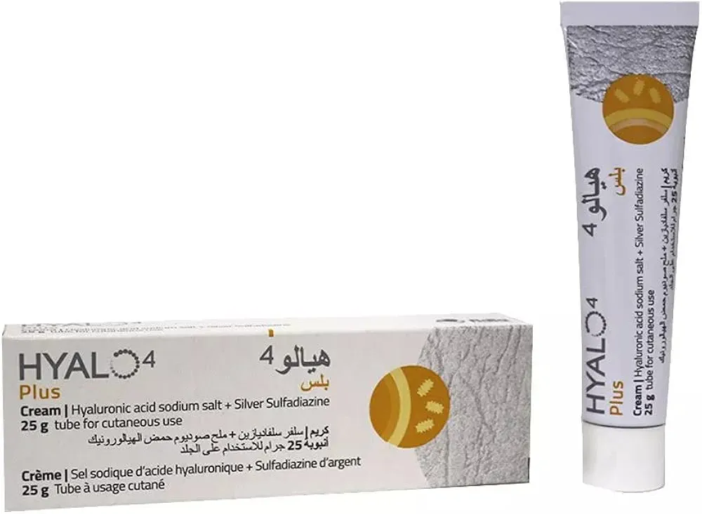
In a modern clinical setting, the management of ankle osteoarthritis requires a multidisciplinary approach that integrates conservative symptom control with advanced surgical intervention. Patients may initially benefit from pharmacological support, including Mediflam D.T / ميديفلام دي تي 50 mg for inflammation, alongside topical therapies like Hyalo4 plus cream / هيالو 4 بلس كريم 0.2% / 1% and Arthri-Flex Cream / كريم أرثري-فليكس Varies by formulation. When conservative measures are exhausted, clinical decision-making is guided by specialized literature, such as Chronic Ankle Arthritis: A Detailed Clinical Case Study & Diagnostic Approach and Total Ankle Arthroplasty for End-Stage Ankle Osteoarthritis in Ballet Dancers: A Comprehensive Review. Surgical planning often involves joint-preserving procedures such as Supramalleolar Opening-Wedge Osteotomy for Varus Ankle Osteoarthritis: A Masterclass, Supramalleolar Osteotomy: An Intraoperative Masterclass for Ankle Deformity Correction, or [جراحة قطع عظم الساق فوق الكاحل لعلاج خشونة مفصل الكاحل بتقنية الوتد المفتوح: دليل شامل مع الأستاذ الدكتور محمد هطيف](https://www.hutaifortho.com/ar/hub/%D8%AC%D8%B1%D8